1909
Evaluation of renal vascular in living donors before transplantation using non-contrast enhanced magnetic resonance angiographyXiaotian Li1, Lihua Chen1, Zhizheng Zhuo2, and Wen Shen1
1Tianjin First Center Hospital, Tianjin, China, 2Philips Healthcare, Beijing, China
Synopsis
Previous studies showed that contrast-enhanced magnetic resonance angiography can be used for preoperative renal transplantation donor vascular evaluation, but requires exogenous contrast agent. Non-contrast-enhanced magnetic resonance angiography (NCE-MRA) can describe vascular anatomy without contrast agent. Thus, the purpose of this study was to evaluate NCE-MRA for the preoperative evaluation of potential living kidney donors.
Target audience
Researchers and clinicians with an interest in assessment of living renal donors with non-invasion and non-contrast agent.Introduction
Kidney transplantation is the best choice for patients with end-stage renal disease. Increasing numbers of living donor kidney transplants have being performed worldwide, and the majority of donor operations are now laparoscopic1.Preoperative imaging of donor renal vascular anatomy is critical for determining eligibility for laparoscopic nephrectomy as well as the kidney chosen for harvest. Factors for determining donor suitability and ease of laparoscopic nephrectomy include the number of renal arteries and renal veins, the presence of early arterial bifurcation or late venous confluence, and incidental findings2. Computed tomographic angiography (CTA) is a reliable technique for the evaluation of potential renal donors, but it exposes healthy donors to radiation and potentially nephrotoxic iodinated contrast agent. Previous studies showed that contrast-enhanced magnetic resonance angiography can be used for preoperative renal transplantation donor vascular evaluation, but requires exogenous contrast agent2,3. Non-contrast enhanced magnetic resonance angiography (NCE-MRA) can describe vascular anatomy without contrast agent. Thus, the purpose of this study was to evaluate NCE-MRA for the preoperative evaluation of potential living kidney donors and compare MR findings with CTA.Methods
Six prospective living renal donors (mean age 40.7±8.09 years, 2 female and 4 male) were prospectively recruited. Prior to NCE-MRA scan, all subjects underwent CTA. NCE-MRA studies were performed on a 3.0T scanner (Philips Ingenia 3.0T, Best, The Netherlands) including arterial imaging and venous imaging. Arterial imaging was applied by 3D B_TFE sequence with following parameter: TR=6.9 ms; TE=3.4ms; FOV=300×120×100mm; Voxel Size=1.5×1.5×2 mm; matrix=200×79; NSA=1; flip angle=27°. Venous imaging was applied by 3D PCA sequence with following parameter: TR=10 ms; TE=6.4 ms; FOV 230×230×70mm; Voxel =1.3×1.3×1.3mm; NSA=3; slices=54; flip angle=10°; PC velocity=15 cm/s. Two radiologists observed renal arteries and veins on original image slice by slice and 3D MIP reconstructed image independently. The image quality of renal vessels was evaluated on the basis of a four-point scale4:1, no diagnostic value (no signal within the vessel); 2, moderate (inhomogeneous signal within the vessel, no sharp vessel border delineation); 3, good (homogeneous signal within the vessel with slight flow artifacts, almost complete and sharp delineation of vessel border); and 4, excellent (completely homogeneous signal within the vessel lumen without flow artifacts, sharp and complete delineation of vessel border). The vascular anatomy or variations of the arterial and venous systems were recorded. The ability of NCE-MRA and CTA to find vasculature anatomical variation was analyzed by using the McNemar x2 test. Kappa statistics were also calculated to quantify the inter-reader variability for assessment of renal vessels using NCE-MRA. Statistical analysis was performed in SPSS 22.0 software (SPSS Inc., Chicago, IL, USA) where p <0.05 indicated significant difference.Results and Discussion
The image quality was categorized as excellent in 83% (5 of 6), and good in 17% (1 of 6) of the patients. Among 12 renal arteries, several variations of vascular were found, including 2 left accessory artery and 1 right proximal arterial branch. Among 12 renal veins, 1 right accessory vein was observed. There is no statistical difference in the ability between NCE-MRA and CTA to find vasculature anatomical variation (x2 =1, p values= 0.5). Excellent inter-reader agreement was found for assessment of renal vessels (kappa value=1.0, p values <0.001).The image quality of NCE-MRA was acceptable, and the detection of renal vasculature anatomical variation was consistent with CTA results, which confirmed the feasibility of using it for preoperative renal donor vascular assessment, and the diagnostic accuracy was high.Conclusion
NCE-MRA was a noninvasive and non-contrast agent tool for evaluation of the renal vasculature and variations with high accuracy. It would be a good modality in preoperative evaluation of living renal donor.Acknowledgements
No acknowledgement found.References
- Fuller T F. Laparoscopic living donor nephrectomy: making optimal use of donors without doing harm. Transplantation, 2014, 98(11):1144.
- Gulati M, Dermendjian H, Gómez A M, et al. 3.0 Tesla magnetic resonance angiography (MRA) for comprehensive renal evaluation of living renal donors: pilot study with computerized tomography angiography (CTA) comparison. Clin Imaging, 2016, 40(3):370-377.
- Kuhlemann J, Blondin D, Grotemeyer D, et al. Gadofosveset-enhanced MR imaging for the preoperative evaluation of potential living kidney donors: correlation with intraoperative findings. Rofo, 2010, 182(11):1001-1009.
- Tang H, Wang Z, Wang L, et al. Depiction of transplant renal vascular anatomy and complications: unenhanced MR angiography by using spatial labeling with multiple inversion pulses. Radiology, 2014, 271(3):879-887.
Figures
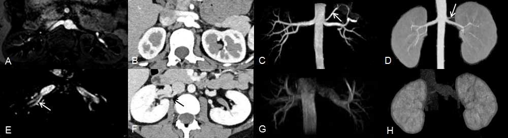
Figure 1. Images in a 34-year-old woman. Original NCE-MRA image and CTA image of renal arteries (A, B); MIP reconstructed NCE-MRA image and CTA image of renal arrteries (B, C) show left proximal arterial branch (arrow);Original NCE-MRA image and CTA image of renal veins(E, F) show right accessory vein (arrow); MIP reconstructed NCE-MRA image and CTA image of renal veins(G, H).