1895
Shortening scan times for functional lung imaging using matrix pencil decomposition1Division of Radiological Physics, Department of Radiology, University of Basel Hospital, Basel, Switzerland, 2Department of Biomedical Engineering, University of Basel, Basel, Switzerland, 3Pediatric Respiratory Medicine, Department of Pediatrics, Inselspital, Bern University Hospital, Bern, Switzerland
Synopsis
This study examines the performance of functional lung MRI using matrix pencil (MP) decomposition for the estimation of regional fractional ventilation and perfusion by exploring the relationship between the scanning time and the quantitative outcomes. Our results show excellent promise that the overall scan time for functional lung imaging using MP decomposition can be considerably shortened, which can allow for easier integration of MP MRI in clinical routine protocols.
Introduction
Recently, matrix pencil (MP) decomposition1 has been proposed as an improvement over Fourier decomposition2 MRI for the regional assessment of the lung function. MP MRI offers better performance than fast Fourier transform based analysis for truncated time-series and has shown to be a rapid and robust tool for the assessment of regional ventilation and perfusion impairment in a range of lung diseases3-5. Typically, 2D time series of ultra-fast steady state free precession (ufSSFP)6 images are acquired for about one minute, resulting in about 160 time-points for subsequent spectral analysis. In this work, we explore the performance of the matrix pencil method for robust assessment of lung function from time series having different lengths and thus overall acquisition times.Methods
MR data acquisition
Measurements were performed on 1.5T (MAGNETOM Aera and Avanto-Fit, Siemens Healthineers, Germany) MR-scanners using a combination of thorax and spine receiver coils. A healthy volunteer and a cystic fibrosis patient were scanned during free-breathing with a time-resolved 2D ufSSFP sequence using the following parameters: field-of-view=450×450mm2, slice thickness=12mm, TE/TR/TA=0.67/1.46/119ms, flip angle α=65º, bandwidth=2056Hz/pixel, matrix=128×128 (interpolated to 256×256), parallel imaging GRAPPA factor=2, 160 coronal images per slice, 3.33 images/s.
Image processing
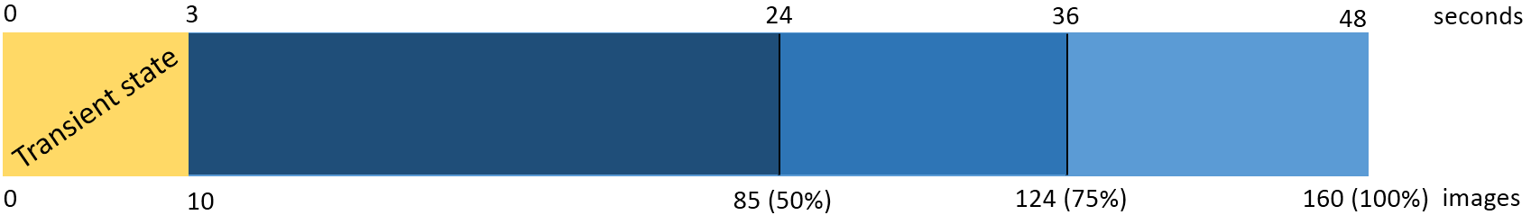
Respiratory motion in the ufSSFP data was compensated with a non-rigid image registration. Subsequently, MP decomposition of the registered and segmented image series was performed voxel-wise to estimate the amplitude of the respiratory and cardiac signal modulations in the lung parenchyma and to create quantitative fractional ventilation (FV) and perfusion (Q) maps2,8. The signal distributions on the segmented FV and Q images were analyzed to estimate threshold values indicating functional impairment equal to 0.75 of median value from the voxel distributions. Eventually, values of relative impaired ventilation (RFV) and perfusion (RQ) were calculated. The performance of MP decomposition was analyzed in post-processed time-series of different length N between 75 to 150 images (cf. Figure 1).
Results
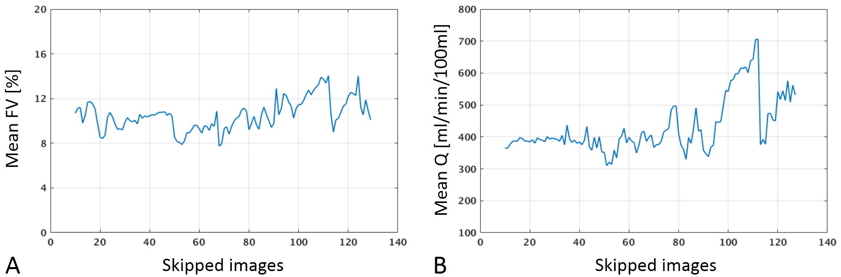
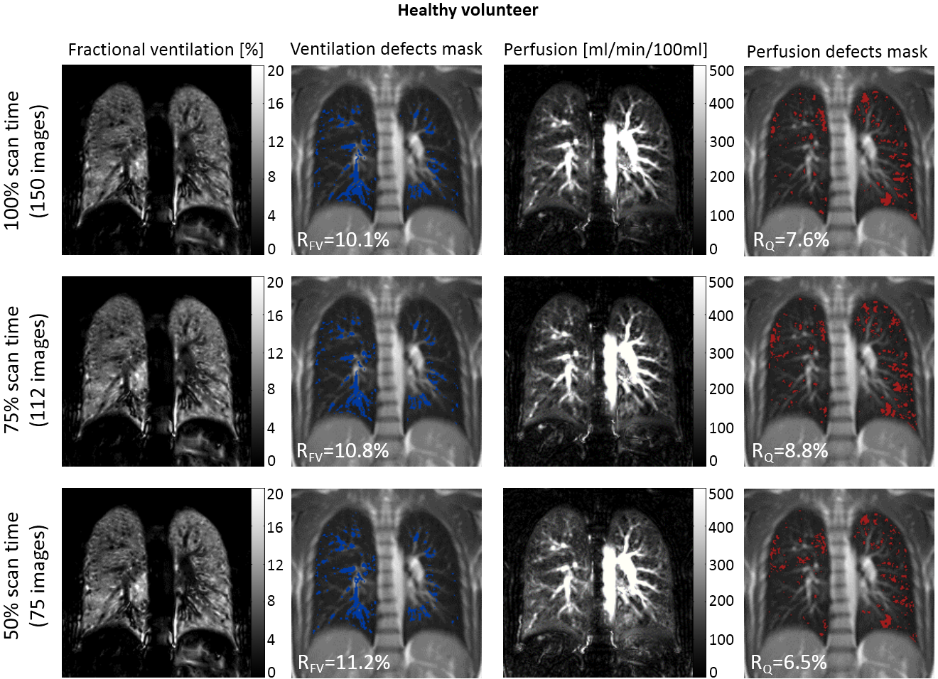
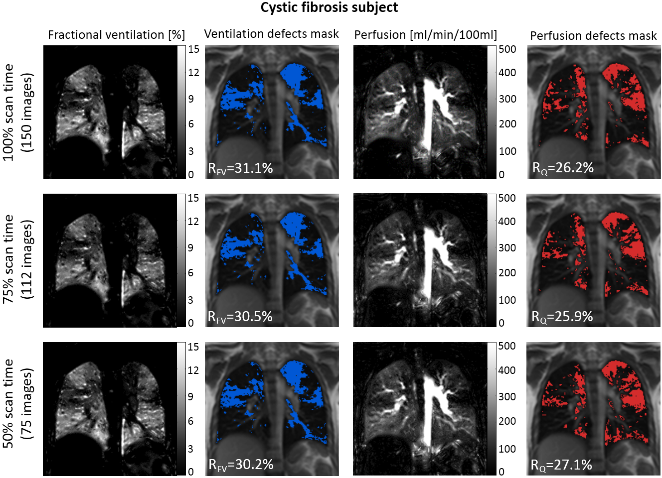
Figure 2 shows mean values of fractional ventilation and perfusion as a function of the length of the time-series measured in a cystic fibrosis patient. It can be noticed that the reduction of the dataset length by more than 50% strongly affects estimated values of fractional ventilation and perfusion. Figure 3 and 4 present exemplary fractional ventilation and perfusion maps as well as masks showing regions with impaired lung function acquired in a healthy volunteer and a cystic fibrosis patient. The images were generated from time-series containing 100%, 75% and 50% of the data (150, 112 and 75 images with corresponding scan-times 48, 36 and 24 seconds). The values of RFV and perfusion RQ remains stable despite the reduction of the length of the dataset.Discussion and Conclusion
For the whole lung functional assessment using MP MRI, typically a series of eight to twelve 2D series of 160 images are acquired; leading to about ten minutes of scan time. Using MP analysis, the time-series can be considerably shortened, without affecting the outcome values of the relative fractional ventilation and perfusion impairment. Our results show excellent promise that the overall scan time for functional lung imaging using MP decomposition can be considerably shortened. This can allow for easier integration of MP MRI in clinical routine protocols. The robustness of the MP approach will be further evaluated in data already acquired in different patient cohorts3-5.
Acknowledgements
References
1. Bauman G, Puderbach M, Deimling M et al. Non-contrast-enhanced perfusion and ventilation assessment of the human lung by means of fourier decomposition in proton MRI. Magn Reson Med. 2009 Sep;62(3):656-64.
2. Bauman G, Bieri O. Matrix pencil decomposition of time-resolved proton MRI for robust and improved assessment of pulmonary ventilation and perfusion. Magn Reson Med. 2017 Jan;77(1):336-342.
3. Nyilas S, Bauman G, Sommer G et al. Novel magnetic resonance technique for functional imaging of cystic fibrosis lung disease. Eur Respir J 2017 Dec 7;50(6.
4. Nyilas S, Bauman G, Pusterla O et al. Ventilation and perfusion assessed by functionalMRI in children with CF: reproducibility in comparison to lung function. J CystFibros. 2018 Oct 19.
5. Nyilas S, Bauman G, Pusterla O et al. Structural and Functional Lung Impairment in PCD: Assessment with MRI and Multiple Breath Washout in Comparison to Spirometry. Ann Am Thorac Soc. 2018 Oct 5.
6. Bauman G, Pusterla O, Bieri O. Ultra-fast steady-state free precession pulse sequence for Fourier decomposition pulmonary MRI. Magn Reson Med. 2016 Apr;75(4):1647-53.
7. Chefd’hotel C, Hermosillo G, Faugeras O. Flows of diffeomorphismsfor multimodal imageregistration. In: Proceedings of the IEEE International Symposium on Biomedical Imaging(ISBI’2002), Washington, DC, USA, July 2002. pp. 753–756.
8. Kjørstad Å, Corteville DM, Fischer A et al. Quantitative lung perfusion evaluation using Fourier decompositionperfusion MRI. Magn Reson Med. 2014 Aug;72(2):558-62.
Figures