1893
In Vivo 19F-Isoflurane Chemical Shift Imaging in mouse lung1Monash Biomedical Imaging, Monash University, Clayton, Australia
Synopsis
Proton MRI of lungs has challenges because of the available signal and high susceptibility of the air and tissue interface. Isoflurane is a commonly used fluorinated anesthetic with an excellent safety record in preclinical studies, which contains small amounts of the stable fluorine isotope 19F. This abstract demonstrates that Isoflurane can be used as both an anesthetic and as 19F contrast agent simultaneously. Inhaled isoflurane provides 19F signals in the gas and the dissolved phases which offers potentials for functional MR imaging of lungs.
Introduction
Lung MR imaging has traditionally been very challenging due to the low density of protons and the high susceptibility of lung tissue, traditional proton MRI techniques cannot provide high-quality pulmonary images. Hyperpolarized gases, such as 3He and 129Xe, can be used as exogenous contrast agents for pulmonary imaging1. However, these gases have limitations due to their high cost, requirement for specialized equipment and rapid depolarization in the presence of oxygen, which all restrict their clinical applications. Unlike hyperpolarized gases, fluorinated agents can be mixed with oxygen and are readily available at a reasonable cost. Perfluoropropane has been used as contrast agent for anatomical pulmonary imaging2, however, perfluoropropane is not soluble in water and cannot provide information on the dissolved-phase in lungs. Isoflurane is a water soluble fluorinated agent and the most commonly used inhaled anesthetic in pre-clinical studies. In this study, we aimed to use isoflurane as the contrast agent to evaluate its potential to provide useful pulmonary imaging information.Materials and Methods
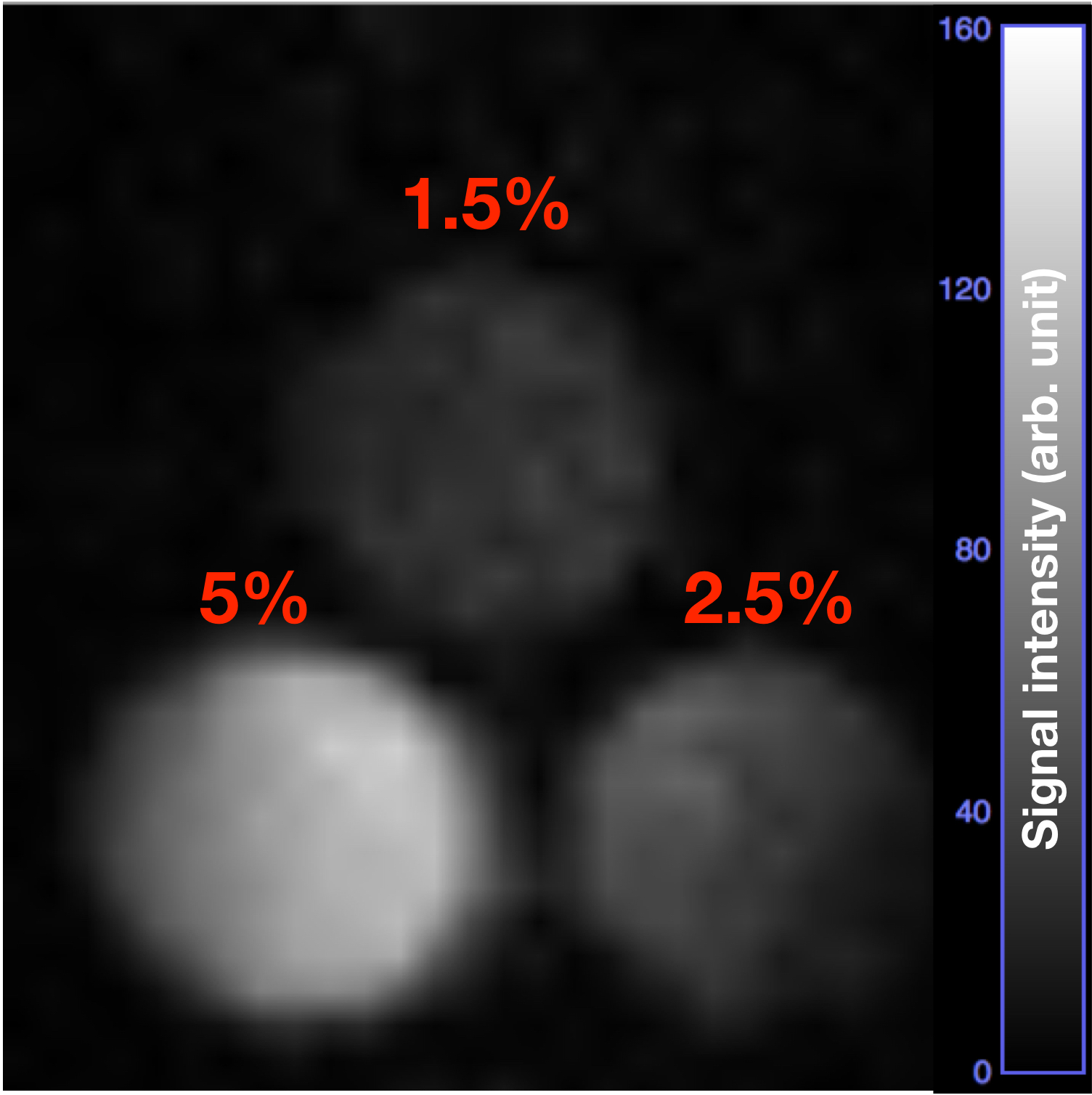
All MRI experiments were performed on a Bruker BioSpec 9.4T scanner (Bruker BioSpin GmbH, Ettlingen, Germany) with a 19F/1H dual-channel 40mm volume coil. A phantom filled with 100% isoflurane liquid (IsoFlo, Zoetis Australia Pty Ltd) was used to detect the two resonant frequencies of 19F. Three gas-phase phantoms with 1.5%, 2.5% and 5% isoflurane were applied to test the gas-phase signals of isoflurane. Two C57Bl6 mice were used in this study and anesthetized with 1-1.5% isoflurane without 19F scans. The isoflurane was increased to 2.5% during the 19F imaging.Data Acquisition
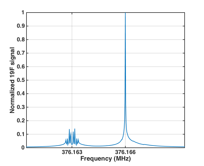
The resonant frequency for 19F chemical shift imaging was set at 376.166 MHz. A 3D RARE sequence was used for imaging 19F-isoflurane. The parameters for imaging the liquid 19F phantom were TR=1000ms, TE=5.17ms, FOV=40*40*20mm3, Rare factor=2, matrix=32*32*8, bandwidth=10000Hz, and axial plane. The parameters for imaging the gas-phase 19F phantoms were TR=1000ms, TE=5.17ms, FOV=40*40*240mm3, Rare factor=2, matrix=32*32*8, bandwidth=10000Hz, and axial plane orientation with 20 averages. The parameters for imaging the gas-phase 19F phantoms were TR=1000ms, TE=5.17ms, FOV=40*40*240mm3, Rare factor=2, matrix=32*32*8, bandwidth=10000Hz, axial plane orientation with 20 averages. The parameters for in-vivo 19F imaging were TR=1000ms, TE=4.6ms, Rare factor=2, matrix=32*32*8, bandwidth=10000 Hz, 2 averages, imaging time=4mins16s, coronal plane with FOV=32*32*160mm3 and axial plane with FOV=32*32*100mm3 in two mice, respectively.Results and Discussion
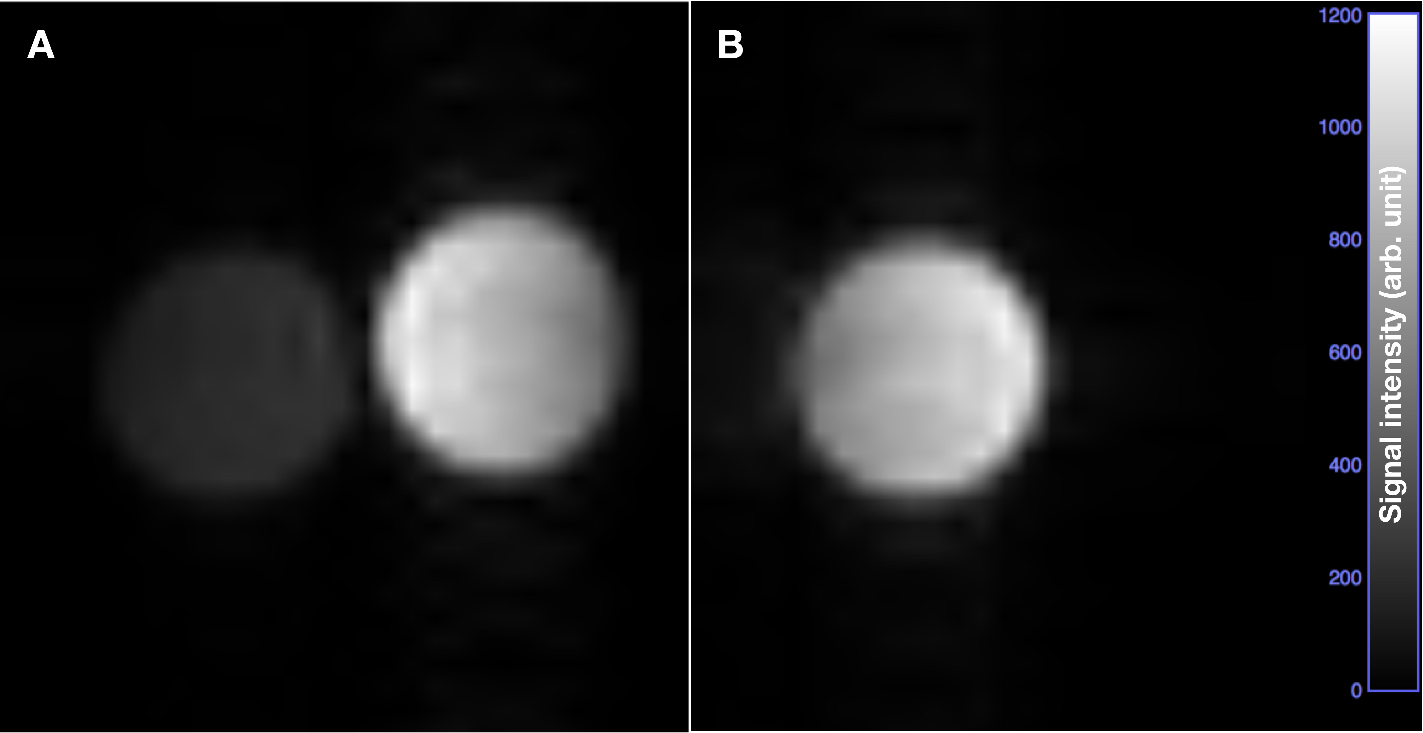
Imaging a liquid isoflurane sample showed two main resonant peaks with frequencies of 376.163 and 376.166 MHz (Figure 1). The normalized signal of the 376.166 MHz peak was much higher, and hence this frequency was used for all 19F imaging. Low proton signals can be seen in 1H-channel image of liquid isoflurane (Figure 2A left), compared with high proton signals in water (Figure 2A right). Robust 19F signal can be detected in the liquid isoflurane phantom using the 19F-channel, while no signal was detected in the water sample (Figure 2B).
To test the utility of measuring the gas-phase isoflurane signal at standard concentrations used for anesthesia, a range of isoflurane concentrations were tested mixed with pure oxygen (Figure 3). The results showed a concentration dependent signal, with 2.5% isoflurane providing a reasonable 19F signal for imaging. Considering both imaging and anesthesia constraints, a short-term experiment testing 19F imaging in live mice was performed with 2.5% isoflurane.
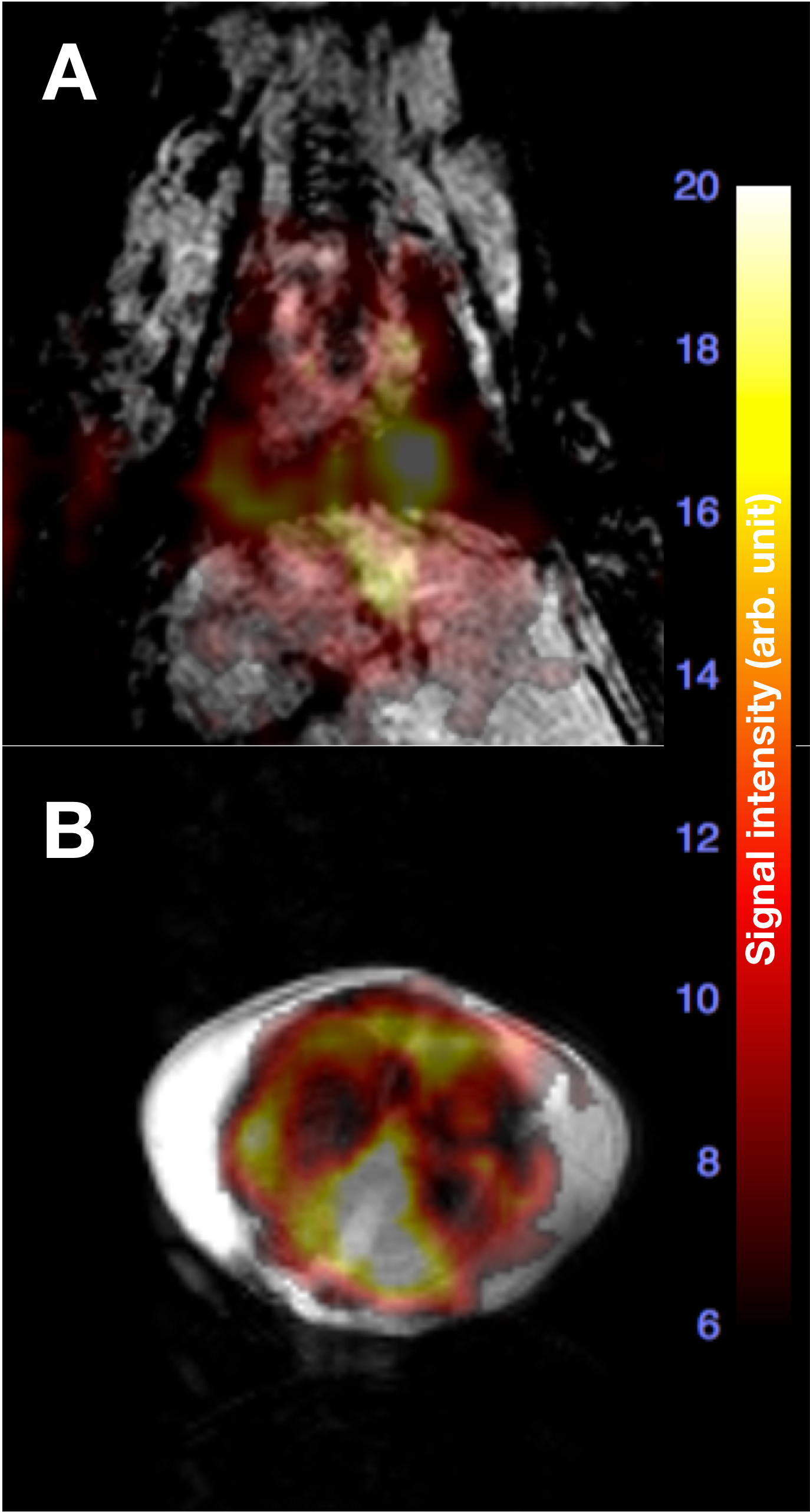
In vivo the 19F signal was mainly accumulated in the lung, liver, and heart, supporting prior data showing that isoflurane accumulated in these organs3(Figure 4A and 4B). It was interesting that isoflurane markedly accumulated in pleura (Figure 4B). The 19F signals in liver (Figure 4A) and heart (Figure 4A and 4B) could be attributed to the uptake of dissolved isoflurane resulting from gas exchange in the lung to the blood. Because the gas-phase of isoflurane can be imaged with the same resonant frequency, the 19F signals in lung could be coming from either the dissolved or gas-phase isoflurane. The concentration of dissolved isoflurane could increase with sustained anesthetic delivery4, which might help to enhance the 19F signals in lung. Further experiments will be required to determine is isoflurane can be used to differentiate dissolved and gas phase lung imaging.
Conclusions
19F-isoflurane can provide both gas and dissolved phase 19F signals in lung and can be used as an inhalational contrast agent for pulmonary imaging.Acknowledgements
We would like to acknowledge the support of the Australian National Imaging Facility Fellowship.References
[1] Roos JE, McAdams HP, Kaushik SS, Driehuys B. Hyperpolarized Gas MR Imaging: Technique and Applications. Magn Reson Imaging Clin N Am. 2015;23(2):217-29.
[2] Halaweish AF, Moon RE, Foster WM, Soher BJ, McAdams HP, MacFall JR, Ainslie MD, MacIntyre NR, Charles HC. Perfluoropropane gas as a magnetic resonance lung imaging contrast agent in humans. Chest. 2013 Oct;144(4):1300-1310.
[3] Kaye AD, Kaye AM, Urman RD. Essentials of pharmacology for anesthesia, pain medicine, and critical care. New York: Springer 2015.
[4] Venkatasubramanian PN, Shen YJ, Wyrwicz AM. Characterization of the cerebral distribution of general anesthetics in vivo by two-dimensional 19F chemical shift imaging. Magn Reson Med. 1996;35(4):626-30.
Figures