1890
Spin echo and gradient echo quantification in oxygen enhanced MRI may yield different results1Medical radiation physics, Translational Medicine, Malmö, Sweden
Synopsis
Pulmonary oxygen enhanced MRI is currently performed with a variety of pulse sequences. This abstract provides evidence that the oxygen enhancement, quantified as the change in longitudinal relaxation rate, ΔR1, assessed with a spin-echo or gradient-echo sequence may yield different values. Indeed, a standard IR-HASTE quantification yielded group mean ΔR1 10% higher than the Snapshot-FLASH quantification in 15 healthy volunteers. Although studies employing different quantification schemes are likely comparable, caution is warranted since the mean ΔR1 quantified with HASTE-type sequences is likely higher.
Introduction
A wide array of methods are currently employed for pulmonary T1 quantification and measurements of quantitative oxygen enhanced MRI, and a recent meta-analysis by Dietrich et al. described the state of the art [1]. Although the meta-analysis established that there was no difference in mean T1 or ΔR1 quantified with HASTE or FLASH-based inversion recovery [1], there is reason to believe otherwise. Perialveolar water such as capillary blood and pulmonary surfactant are likely long-T1 and high ΔR1 compartments. However, perialveolar water is also must attenuated by susceptibility gradients, potentially lowering both T1 and ΔR1 in a gradient echo. We present measurements of T1 and ΔR1 in 15 healthy volunteers, quantified with both HASTE and Snapshot-FLASH.Methods
Subjects were prospectively recruited subjectively healthy non-smokers, between 22 to 34 years, consisting of 8 males and 8 females.
All MRI measurements were made on a 1.5 Tesla Siemens Magnetom AvantoFit (Siemens Healthcare, Erlangen, Germany), with an 18 channel body coil and a 32 channel spine matrix.
The Snapshot FLASH protocol was executed with imaging matrix 128 × 64 zero filled to 256 × 256; field of view 450 mm square; slice thickness 1.5 cm; echo time = 0.67 ms; repetition time = 3.0 ms; flip angle = 7°; and 16 images for the T1-quantifiaction.
THE IR-HASTE images were acquired with a resolution of 126x68, interpolated to 256 square; FoV 450 mm square; slice thickness 1 cm; TE = 19 ms; using a global inversion pulse. A complete set of 7 inversion times between 100-3500 was collected for T1 calculation.
All measurements were preceded by instructions for breath-hold after a tidal inspiration and subjects were given 10 seconds of free breathing between breath hold. T1 measurements were performed before and after 5 min 100% oxygen breathing with a tight fitting face mask. After pixel-wise T1 calculation in a single central slice, the mean T1 is used.
Results
One subject generated a large negative OE-effect and was thus excluded from the entire analysis, leaving 15 subjects with both T1 and ΔR1 measurements.
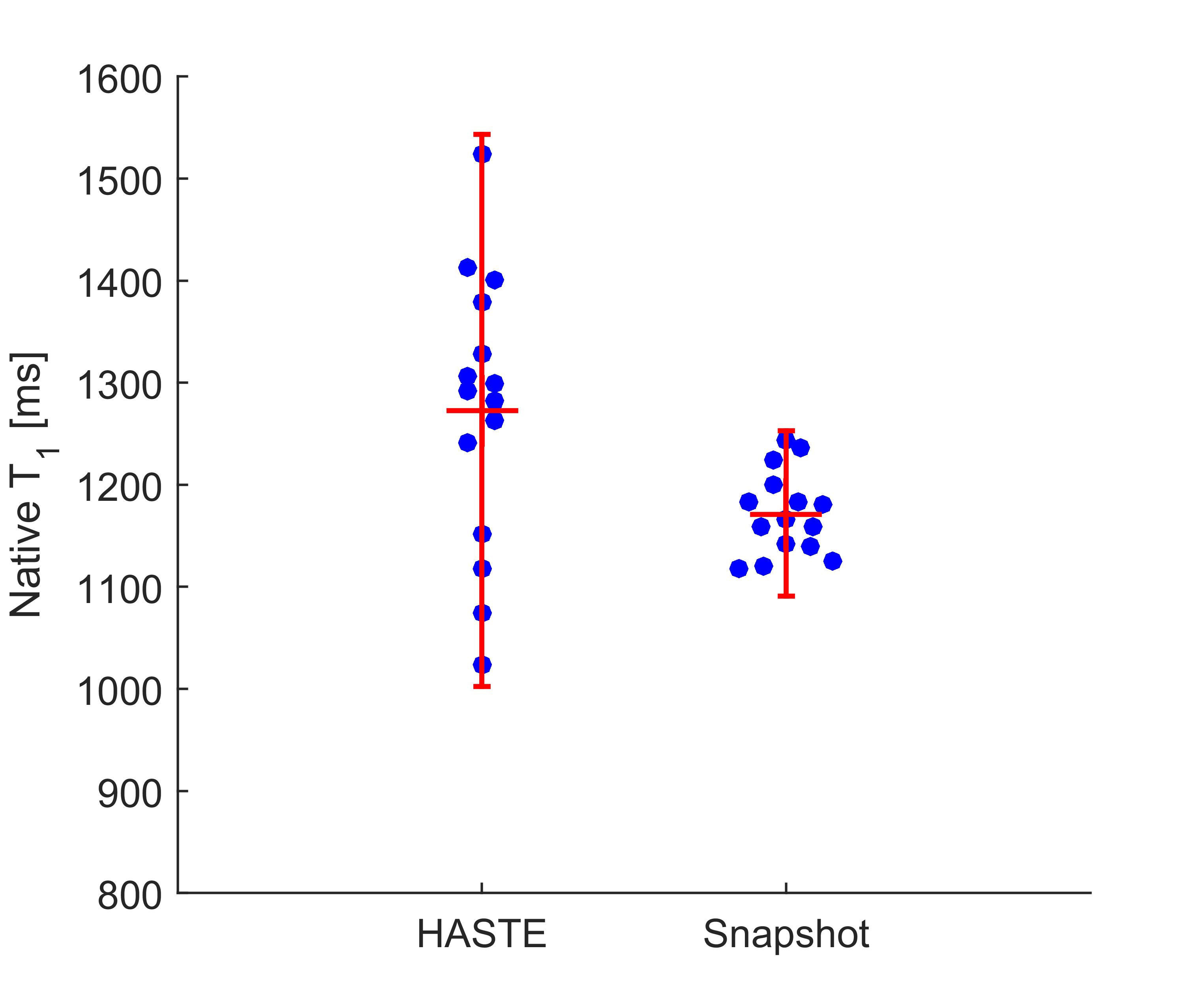
The T1 values determined with both methods are compared in FIGURE 1. The mean T1 is 1290 ms (95%CI [1221, 1360] ms) for the HASTE and 1171 ms (95%CI [1145, 1195] ms) for the FLASH. The 95% CI for the difference in means between the methods is [31, 170] ms at p=0.0076, where the HASTE yield significantly higher values for T1.
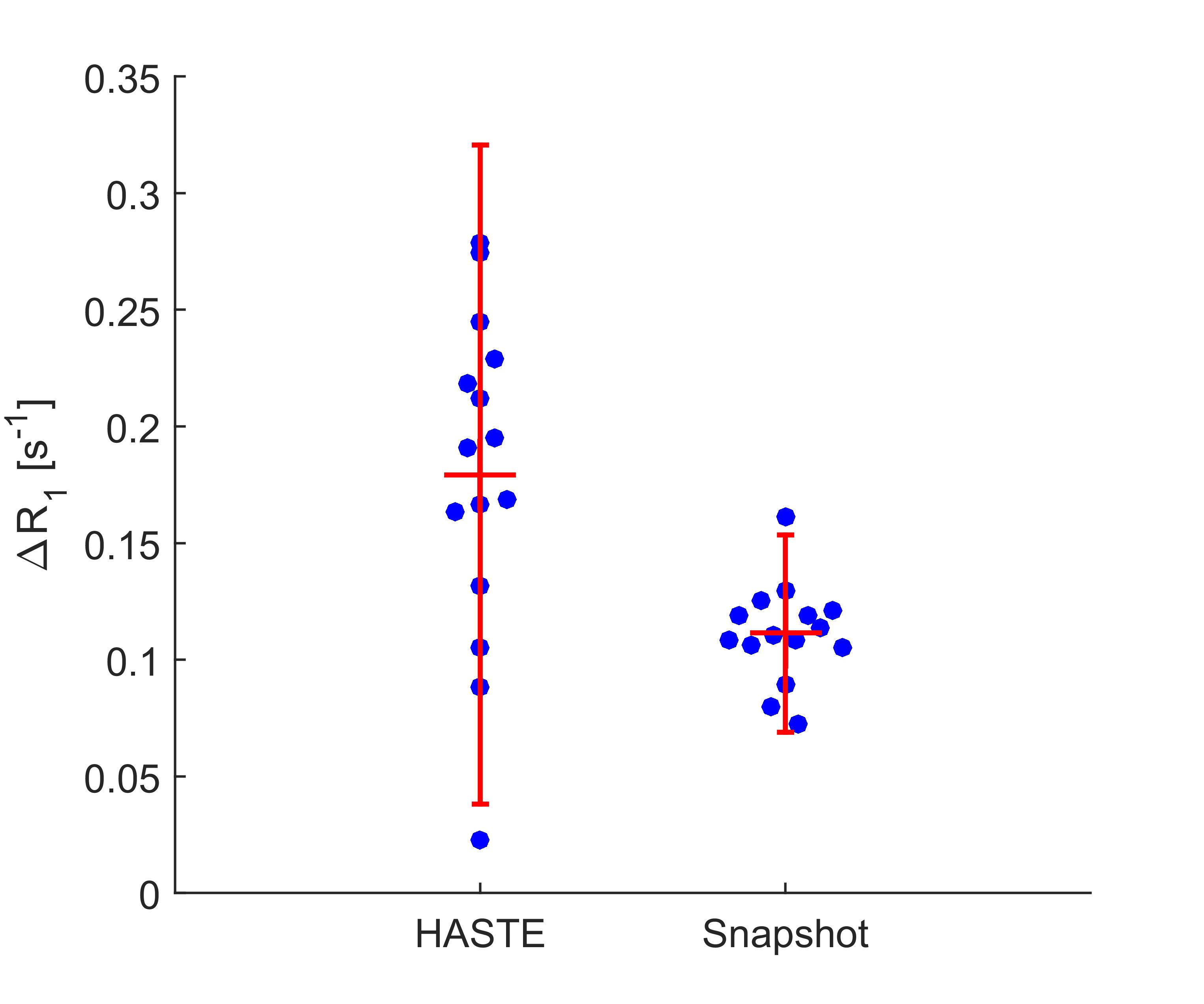
The quantified ΔR1 are presented and compared in FIGURE 2. The mean ΔR1 is 0.180 s-1 (95%CI [0.14, 0.22] s-1) for the HASTE and 0.111 s-1 (95%CI [0.10, 0.12] s-1) for the Snapshot. The HASTE yield significantly higher values for ΔR1 where the 95% CI of the difference in means is [0.03, 0.11] s-1 at p=0.002.
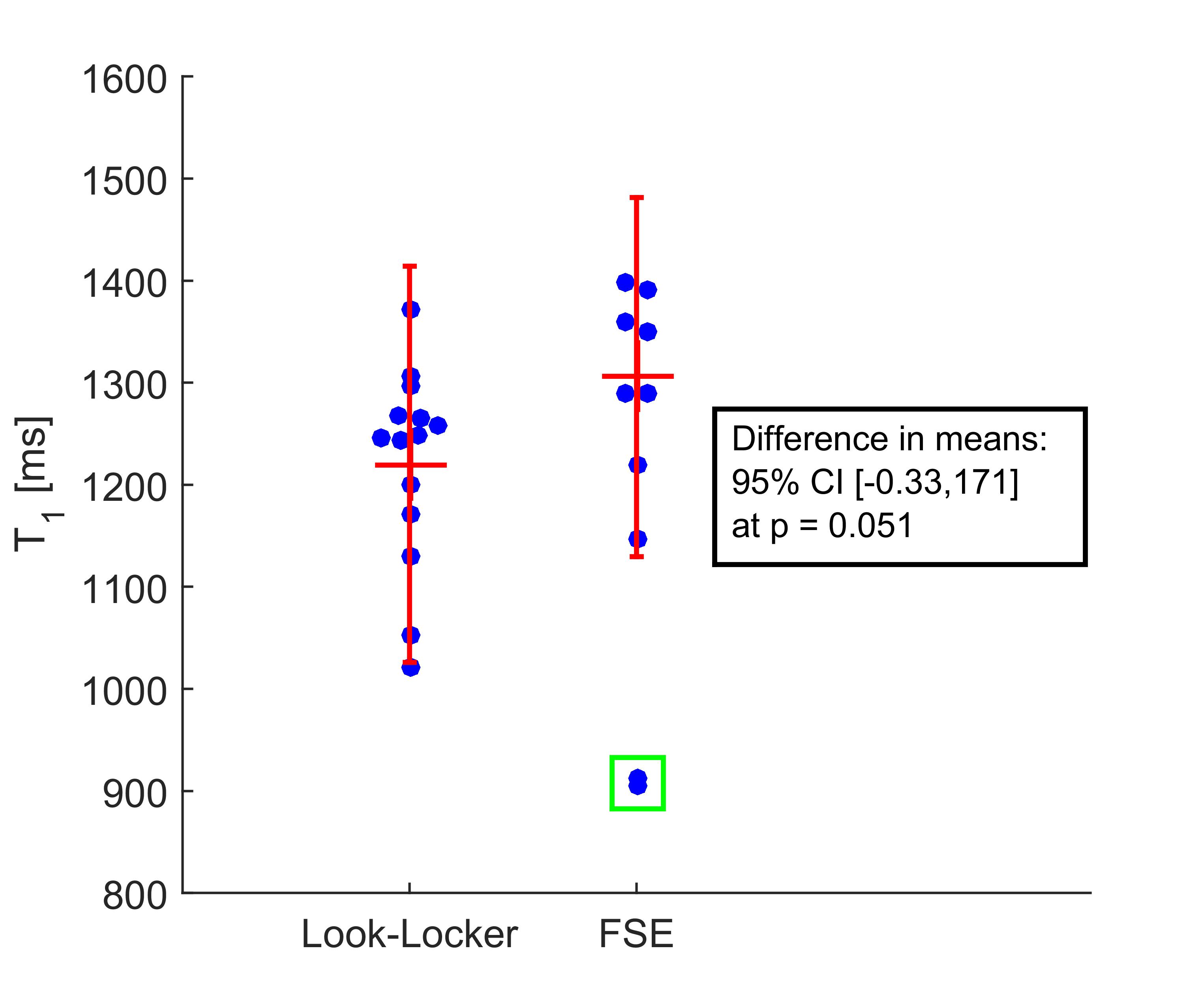
Revisiting the meta-analysis by Dietrich support this data: Indeed, two spin-echo T1 measurements from the meta-analysis are excessively low (900 ms) and are likely generated with slice selective inversion pulses, known to decrease T1 by a substantial amount due to inflow effects [2]. By excluding those two measurements and including the data from the present abstract the 95% CI for difference in means, is [-0.33, 171] ms at p=0.051, suggesting a longer T1 obtained with HASTE (FIGURE 3).
Discussion
It should not be forgotten that measurements with the snapshot FLASH tends to underestimate T1 if anything [3]. However, that HASTE and FLASH would yield different ΔR1 is more controversial. A starting point to investigate this further is the perialveolar water pools. The water closest to the alveolar airspace will have the highest levels of oxygenation and the highest susceptibility gradients during OE imaging, and include pulmonary surfactant and capillary blood.
The results presented in this abstract indicate that the oxygen enhancement effect, as ΔR1, quantified with gradient or spin echoes may be different. This may have a physiological and physical explanation. Although the effect seem to be limited in size to 10% of native T1 values, it warrants some caution when comparing measurements from different centers, using different methods.
Acknowledgements
No acknowledgement found.References
1. Dietrich O, Gaass T, Reiser MF. T1 relaxation time constants, influence of oxygen, and the oxygen transfer function of the human lung at 1.5 T—A meta-analysis. Eur J Radiol. Elsevier Ireland Ltd; 2017;86: 252–260. doi:10.1016/j.ejrad.2016.11.027
2. Wang T, Schultz G, Hebestreit H, Hebestreit A, Hahn D, Jakob PM. Quantitative perfusion mapping of the human lung using 1H spin labeling. J Magn Reson Imaging. 2003;18: 260–5. doi:10.1002/jmri.10338
3. Deichmann R, Haase A, Hubland A. Quantification of Tl Values by SNAPSHOT-FLASH NMR Imaging. J Magn Reson. 1992;96: 608–612.
Figures