1886
Jacobian-based Ventilation Derived from Lung Proton MRI: Correlation with Ventilation Defects on Hyperpolarized Gas MRI1Medical Physics, University of Wisconsin-Madison, Madison, WI, United States, 2Biomedical Engineering, University of Wisconsin-Madison, Madison, WI, United States, 3Biostatistics and Medical Informatics, University of Wisconsin-Madison, Madison, WI, United States, 4Radiology, University of Wisconsin-Madison, Madison, WI, United States, 5Pharmacy, University of Wisconsin-Madison, Madison, WI, United States, 6Allergy, Pulmonary, & Critical Care Medicine, University of Wisconsin-Madison, Madison, WI, United States
Synopsis
We used deformable registration between 1H MRI at two lung volumes to create a Jacobian based ventilation map for quantification of ventilation heterogeneity. We compared values derived from the Jacobian ventilation map to the ventilation defect percent (VDP) from hyperpolarized Helium-3 MRI (HP 3He MRI) and to spirometry measures and found that the Jacobian minimum was associated with both VDP and FEV1/FVC %. These findings suggest that ventilation quantified using Jacobian maps of deformable registration on 1H MRI is a potential alternative to HP MRI ventilation imaging.
Introduction
Hyperpolarized (HP) 3He MRI derived ventilation measures, such as VDP, have previously been established as a predictor for severe clinical outcomes in asthma1,2. However, the time and expense of conducting HP MRI has thus far limited its possible use in a clinical setting. Jacobian ventilation maps have been used to estimate ventilation from 4DCT3,4,5. Prior work has used registered 1H lung MRI in conjunction with signal differences to detect local ventilation abnormalities6. In this work, we present a method to create Jacobian ventilation maps from 1H MRI and compare measurements derived from these maps to VDP derived from HP 3He MRI in the same subjects. We hypothesized that reduced deformation (low Jacobian values) would be more specifically associated with the regional obstruction measured by the VDP.Methods
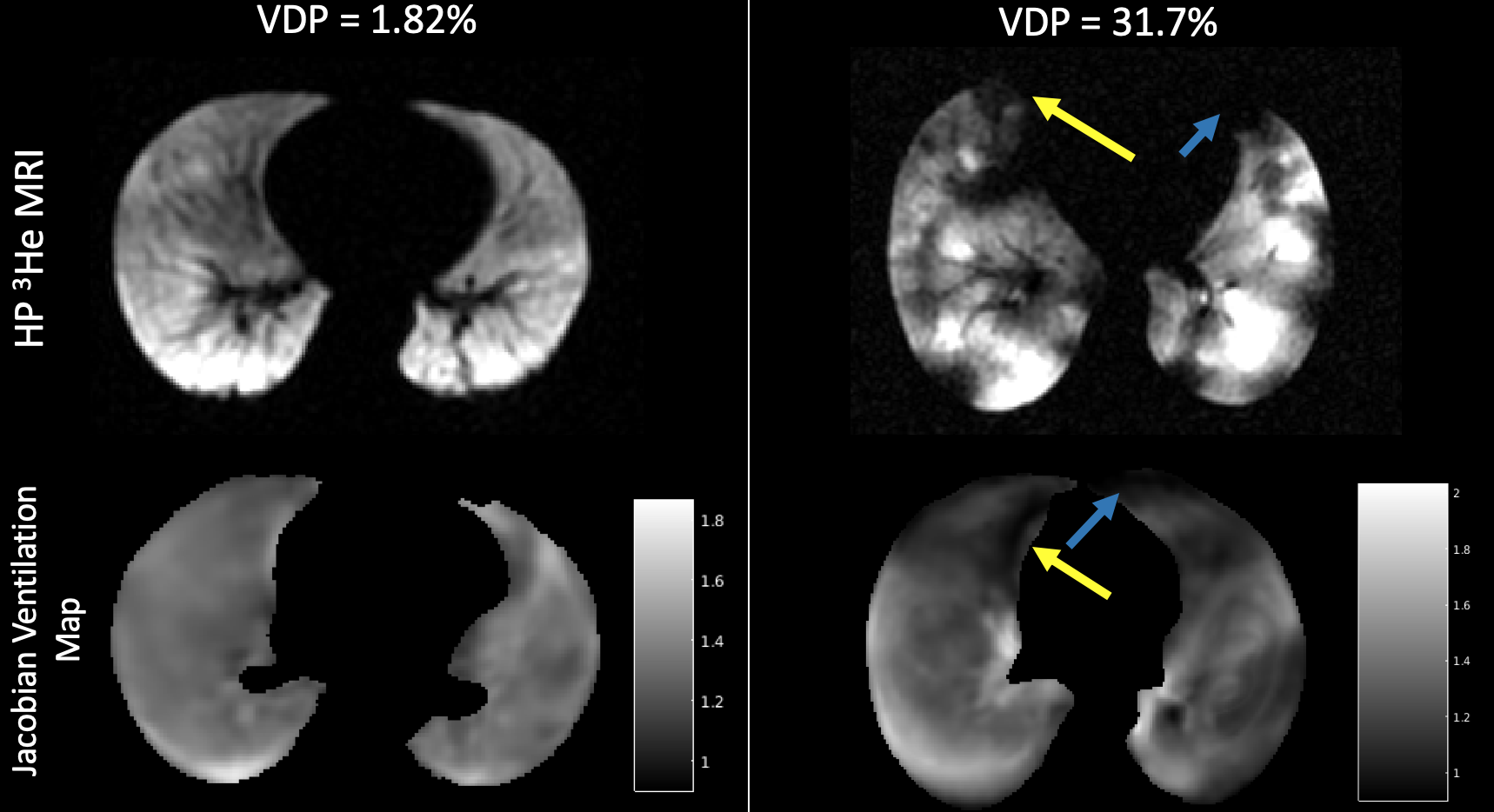
Our population was drawn from the Severe Asthma Research Program III (SARP3) and consisted of 29 asthmatics (8 mild/moderate, 21 severe; 13M 16F; age 41.3 ±19.4 yrs). Proton and HP 3He MRI were obtained using a 1.5T MRI (GE Healthcare, Waukesha, WI) and spirometry was obtained on the same visit. HP 3He MRI was performed during a breath-hold from functional residual capacity (FRC) using a volume of gas estimated to be 14% of the subject’s total lung capacity (TLC) (~FRC+1L). Proton MRI included breath held acquisitions obtained at FRC and at a volume matching 3He MRI using a 3D SPGR sequence. Parameters for both sequences are shown in Table 1. The whole lung ventilation defect percent (VDP) was calculated from HP 3He MRI7. As summarized in Figure 1, the 1H SPGR acquired at the 3He-matched lung volume was registered to the 1H SPGR acquired at FRC lung volume using a B-Spline deformable registration8. The voxel wise Jacobian map is the determinant of the deformation gradient tensor calculated from the registration deformation field between the two different lung inflation volumes, providing direct measures of regional volume changes. The Jacobian minimum, or the minimum value of the Jacobian map within the lungs, was calculated for each subject. Spearman’s correlation was used to compare Jacobian-derived metrics with VDP and spirometry.Results
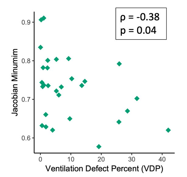
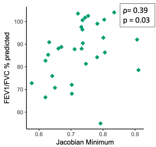
Areas of low values on the Jacobian map were qualitatively associated with areas of ventilation defect on HP 3He (Figure 2). The Jacobian minimum was associated with VDP from HP 3He MRI (ρ= -0.38, p = 0.04) (Figure 3) as well as FEV1/FVC percent predicted (ρ= 0.39, p = 0.03) (Figure 4). The Jacobian minimum was not associated with FEV1 or FVC percent predicted.Discussion
The Jacobian minimum is associated with the well-established VDP values, which have been previously shown to predict asthma exacerbations in this population1,2. The Jacobian minimum was also associated with the FEV1/FVC percent predicted, which has been previously shown to relate to airflow limitation caused by narrowing of the airways8.These results show that Jacobian ventilation maps created using proton MRI may provide an alternative to HP gas MRI as a means of estimating regional ventilation in asthma.Conclusions
Jacobian based ventilation derived from 1H MRI is associated with VDP and spirometry. Future studies will extend this method to other populations and test its implementation using alternative pulse sequences.Acknowledgements
The authors would like to acknowledge funding from RO1 HL115118 and U10 HL109168, as well as funding from GE Healthcare for MRI research at UW-Madison.References
1. D. G. Mummy et al., “Ventilation defect percent in helium-3 magnetic resonance imaging as a biomarker of severe outcomes in asthma,” J. Allergy Clin. Immunol., vol. 141, no. 3, pp. 1140-1141. e4, 2018.
2. D. Mummy, “High Ventilation Percent on Hyperpolarized Helium-3 MRI is Associated with Reduced One-Year Risk of Asthma Exacerbation,” ISMRM 2018, vol. Abstract 4463.
3. J. M. Reinhardt, K. Ding, K. Cao, G. E. Christensen, E. A. Hoffman, and S. V. Bodas, “Registration-based estimates of local lung tissue expansion compared to xenon CT measures of specific ventilation,” Med. Image Anal., vol. 12, no. 6, pp. 752–763, Dec. 2008.
4. B. A. Tahir et al., “Spatial Comparison of CT-Based Surrogates of Lung Ventilation With Hyperpolarized Helium-3 and Xenon-129 Gas MRI in Patients Undergoing Radiation Therapy,” Int. J. Radiat. Oncol. Biol. Phys., vol. 102, no. 4, pp. 1276–1286, Nov. 2018.
5. T. J. Patton, S. E. Gerard, W. Shao, G. E. Christensen, J. M. Reinhardt, and J. E. Bayouth, “Quantifying ventilation change due to radiation therapy using 4DCT Jacobian calculations,” Med. Phys., vol. 0, no. 0.
6. F. Pennati, J. D. Quirk, D. A. Yablonskiy, M. Castro, A. Aliverti, and J. C. Woods, “Assessment of regional lung function with multivolume (1)H MR imaging in health and obstructive lung disease: comparison with (3)He MR imaging,” Radiology, vol. 273, no. 2, pp. 580–590, Nov. 2014.
7. W. Zha et al., “Regional Heterogeneity of Lobar Ventilation in Asthma Using Hyperpolarized Helium-3 MRI,” Acad. Radiol., 2017.
8. N. J. Tustison and B. B. Avants, “Explicit B-spline regularization in diffeomorphic image registration,” Front. Neuroinformatics, vol. 7, 2013.
9. R. L. Sorkness et al., “Lung function in adults with stable but severe asthma: air trapping and incomplete reversal of obstruction with bronchodilation,” J. Appl. Physiol., vol. 104, no. 2, pp. 394–403, 2008.
Figures
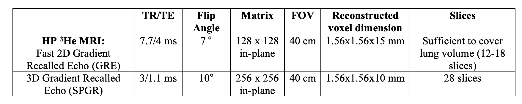
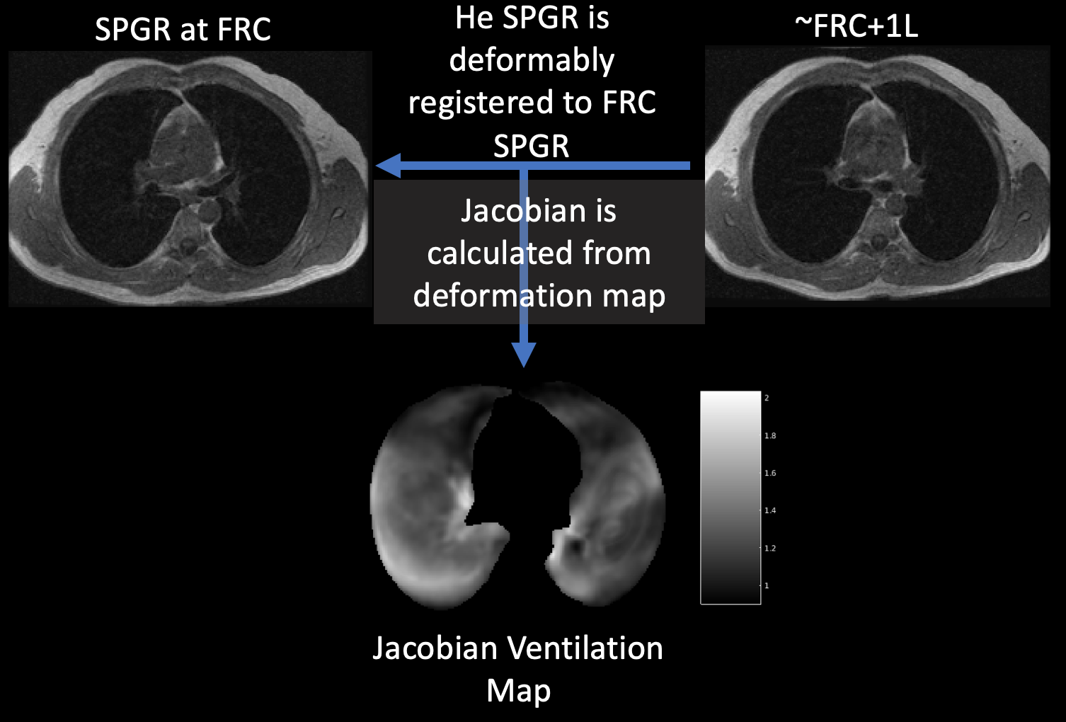
Figure 1: The image analysis pipeline for creating the Jacobian Ventilation map.