1878
Validation of theoretical models of hyperpolarized gas diffusion MRI with finite element simulation in geometrical and realistic models of lung acinar airways from micro-CT1Academic Unit of Radiology, University of Sheffield, Sheffield, United Kingdom
Synopsis
In this work, the stretched exponential (SEM) and cylindrical airway (CM) theoretical models for hyperpolarized gas diffusion MRI are validated with finite element (FE) simulations in two different acinar airway geometries. Simulations of 3He multiple b-value diffusion experiments were performed in a theoretical cylindrical acinar airway geometry and a micro-CT derived realistic acinus model. The simulated MRI diffusion signal was fitted to the CM and SEM. In FE simulations of both models, derived acinar airway parameters from CM and SEM demonstrated strong correlation and good agreement with the underlying model.
Introduction
Theoretical models of hyperpolarized gas diffusion within the lungs, such as the cylinder airway model (CM) and stretched exponential model (SEM), can be used to estimate in-vivo acinar airway dimensions 1,2. Recent work has demonstrated that, across a range of acinar length scales, the SEM mean diffusive length scale (LmD) is non-linearly correlated to the CM-derived mean linear intercept length (LmCM) and is analogous to the CM mean alveolar diameter (LCM) 3. However, unlike the LmCM 1, the LmD parameter has not yet been validated against histology or known geometries. In this work we aim to validate the SEM LmD, alongside CM-derived parameters, with finite element (FE) simulations of hyperpolarized gas diffusion MR in various models of acinar airway geometry.Methods
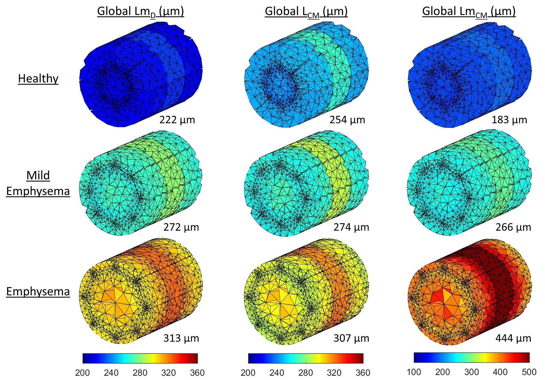
Two different models of acinar airway geometry were designed and meshed for FE simulations that numerically solved the Bloch-Torrey equation describing the hyperpolarized gas MR signal in a pulse gradient diffusion MR experiment. The first type of model was a geometrical model based on the cylindrical acinar airway geometry 1, with seven alveolar sleeve rings, each containing eight alveolus units, surrounding a long alveolar duct (Figure 1a). The outer radius (R), alveolar sleeve depth (h), and mean alveolar diameter (L) were modified to reflect realistic “healthy”, “mild emphysema” and “severe emphysema” acinar dimensions (Table 1). To minimize the effects of finite airway length, the simulation results from the central three rings were considered only 4. The second model was a realistic complete acinar geometry derived from micro-CT segmentation data of an excised sample of healthy human lung 5 (Figure 1b).
A 3He multiple b-value diffusion-weighted experiment (Δ=1.6 ms, b-values=[0, 1.6, 3.2, 4.8, 6.4, 8.0] s/cm2) was simulated using COMSOL Multiphysics for each FE model. The diffusion gradient parameters and the resultant diffusion regimes are consistent with those reported for reliable range of operation of the CM 4,6. Diffusion simulations were solved for 91 and 62 different diffusion-sensitizing gradient angles uniformly orientated in 3D space for the geometrical and realistic acinus models, respectively 4. For every mesh node, the simulated MR signal for each b-value was averaged over all simulated orientations, and fitted to the CM 1 and SEM 2 in MATLAB. For the cylindrical airway geometrical models, the diffusion model-derived estimates of acinar dimension were validated against the actual defined model dimensions; while for the realistic acinar geometry, diffusion-derived parameters were compared to the acinus Lm calculated theoretically (Lm=4V/S) and from direct measurement of linear intercept lengths (Figure 1c).
Results and Discussion
A summary of geometrical model parameters, and diffusion model estimates of acinar dimension for each simulation are shown in Table 1. Distributions of SEM-derived LmD, and CM-derived LCM and LmCM for the three cylindrical acinar airway geometries and the realistic acinus geometry are shown in Figure 2 and 3, respectively. Across the three cylindrical airway geometries, LmD demonstrates good agreement with LCM, and is non-linearly correlated with LmCM, matching the trends observed in recent in-vivo comparisons 3. MR diffusion simulation derived CM parameters were strongly correlated with the actual cylindrical geometry dimensions (r=0.979, P<0.001) and lie near the line of equality (Figure 4). This was expected as the CM diffusion model was optimized for a cylindrical acinar geometry. Reasonable agreement was confirmed with Bland-Altman analysis where a mean bias of 6.5 µm difference in acinar airway parameter was obtained (Figure 4); The observed differences in Lm were similar to those between 3He diffusion Lm and histologically-derived Lm obtained from explanted human lungs 1. Between LmD and LCM, the LmD parameter demonstrated the closest agreement with the cylindrical geometry defined mean alveolar diameter (L), with a mean difference of 5.7 µm across the three cylindrical geometries.
In the realistic acinus geometry, simulation derived LmD, LCM, and LmCM demonstrated good agreement with both theoretical and direct measurements of Lm. The discrepancy between the theoretical and direct Lm could be associated with inherent small differences in the two Lm methodologies 7. Nevertheless, in a healthy acinus model, the LmD value from a simulated 3He Δ=1.6 ms experiment shows good agreement with Lm measurements. Further FE simulations are required at different diffusion times or b-values and with 129Xe gas to fully evaluate the operational range of the SEM and LmD.
Conclusion
This work demonstrates the first validation of the SEM-derived LmD with FE simulations of 3He gas diffusion in cylindrical acinar airway and realistic acinus geometries. SEM and CM-derived parameters both showed good agreement with geometrical model dimensions and mean linear intercept length measurements.Acknowledgements
This work was supported by NIHR grant NIHR-RP-R3-12-027 and MRC grant MR/M008894/1. The views expressed in this work are those of the author(s) and not necessarily those of the NHS, the National Institute for Health Research or the Department of Health.References
1. D. A. Yablonskiy, A. L. Sukstanskii, J. C. Woods, D. S. Gierada, J. D. Quirk, J. C. Hogg, et al., "Quantification of lung microstructure with hyperpolarized 3He diffusion MRI," J Appl Physiol (1985), vol. 107, pp. 1258-65, Oct 2009.
2. H. F. Chan, N. J. Stewart, J. Parra-Robles, G. J. Collier, and J. M. Wild, "Whole lung morphometry with 3D multiple b-value hyperpolarized gas MRI and compressed sensing," Magn Reson Med, vol. 77, pp. 1916-1925, May 2017.
3. H. F. Chan, G. J. Collier, N. D. Weatherley, and J. M. Wild, "Comparison of in vivo lung morphometry models from 3D multiple b-value 3He and 129Xe diffusion-weighted MRI," Magn Reson Med, 2018. DOI:10.1002/mrm.27608.
4. J. Parra-Robles and J. M. Wild, "The influence of lung airways branching structure and diffusion time on measurements and models of short-range 3He gas MR diffusion," J Magn Reson, vol. 225, pp. 102-13, Dec 2012.
5. J. Parra-Robles, B. Veeckmans, M. Rao, J. C. Hogg, and J. M. Wild, "Hyperpolarized gas MR diffusion simulations and experiments in realistic 3D models and phantoms of human acinar airways," in Proc Intl Soc Mag Reson Med, 2015, p. 0160.
6. A. L. Sukstanskii, M. S. Conradi, and D. A. Yablonskiy, "(3)He lung morphometry technique: accuracy analysis and pulse sequence optimization," J Magn Reson, vol. 207, pp. 234-41, Dec 2010.
7. L. Knudsen, E. R. Weibel, H. J. Gundersen, F. V. Weinstein, and M. Ochs, "Assessment of air space size characteristics by intercept (chord) measurement: an accurate and efficient stereological approach," J Appl Physiol (1985), vol. 108, pp. 412-21, Feb 2010.
Figures