1873
Distortion correction in diffusion-weighted MRI of the breast: using point spread function (PSF) encoding1Center for Biomedical Imaging Research, Department of Biomedical Engineering, School of Medicine, Tsinghua University, Beijing, China, 2Philips Healthcare, Beijing, China
Synopsis
Diffusion-weighted MRI of the breast is a promising technique that can detect and characterize breast cancer sensitively and safely. However, geometric distortions of echo-planar imaging (EPI) due to field inhomogeneity and eddy currents has limited its clinical application. Conventional correction methods in breast DWI need additional B0 field maps that are either measured or estimated, and achieve limited effects at tissue interfaces. In this work, we attempted the feasibility of the distortion-free breast DWI using a point spread function (PSF) encoded method. The results showed higher fidelity and better image quality on breast DWI, which could facilitate the anatomical/DWI image registration and improve the quantitative measurements of the breast.
Introduction
Breast DWI has shown great potential for the detection and characterization of breast cancer without the need for contrast agents. There is promising evidence on the utility of apparent diffusion coefficient (ADC) values to differentiate benign and malignant breast tumors 1.
The majority of DWI acquisitions in the breast uses the conventional single-shot EPI (SS-EPI) approach. However, the DWI images are plagued by EPI-related artifacts such as geometric distortion, signal dropout and image blurring, due to the magnetic susceptibility changes, especially around the nipple or breast/skin interface 2. Spatial distortions can cause ADC inaccuracies due to inaccurate registration within the DWI sequence 1. Besides, it can also hamper the integration of DWI data with less distorted e.g. DCE-MRI data for multi-parametric analysis of the breast.
Conventional solutions to the problem of distortion in breast DWI fall into the post-processing correction techniques using the B0 map, which is either measured or estimated 3. However, these methods are not sophisticated enough to completely correct the distortions in breast DWI, especially at the interface between aqueous and adipose substances, where the off resonance effects are severe.
With an additional phase-encoding dimension, point spread function encoded EPI (PSF-EPI) 4-12 has been developed to achieve distortion-free and blurring-free DWI straightforwardly. In this study, we aimed to obtain distortion-free breast DWI using the advanced PSF-EPI technique.
Methods
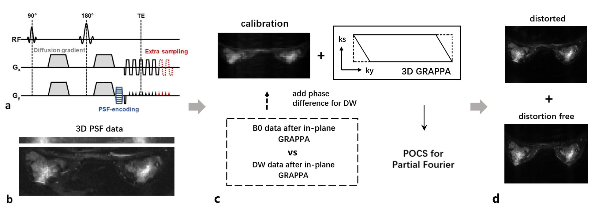
Theory: As demonstrated in Figure 1, PSF-EPI 5 introduces an additional phase-encoding (PE) dimension, called PSF-encoding (kpsf), which is a spin-warp gradient added to the PE direction shot to shot before EPI readout. Integration of the 3D PSF data along the PE (ky) dimension yields a distortion-free image with no chemical shift artifacts, which are otherwise obvious in the PE direction in conventional EPI.
Reconstruction: This work employed a recently developed algorithm named titled-CAIPI 4 for the high acceleration of PSF-EPI. It used a tilted 3D GRAPPA kernel to fill the highly under-sampled k-space, as shown in Figure 1. Projections onto Convex Sets (POCS) was applied for partial Fourier reconstruction along both PE and PSF directions. The shot-to-shot phase corruption due to physiological motion in DWI was corrected via a self-navigated method, which compared the estimated phase of b=0 and diffusion-weighted images from 2D low-frequency kx-ky signals.
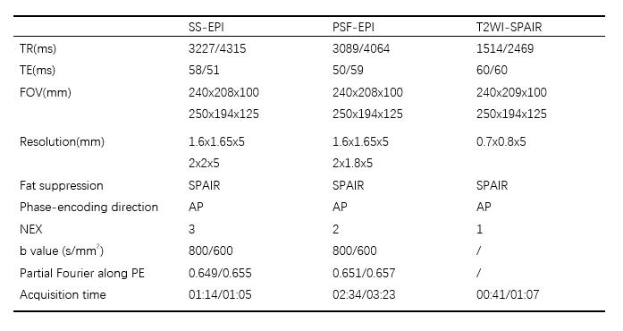
Experiments: Breast MRI was performed on a 3.0T Philips Achieva TX scanner (Philips Healthcare, Best, The Netherlands) with a 7-channel breast coil, including T2-wighted imaging, SS-EPI DWI and PSF-EPI DWI with fat suppression. PSF-related parameters: acceleration factor in PE/PSF direction = 2/11, partial Fourier along PSF =0.545, resulting in 6 shots for signal sampling. A summary of the imaging parameters for breast MRI was listed in Table 1. This study was approved by the Institutional Review Board and written informed consent was obtained.
Results
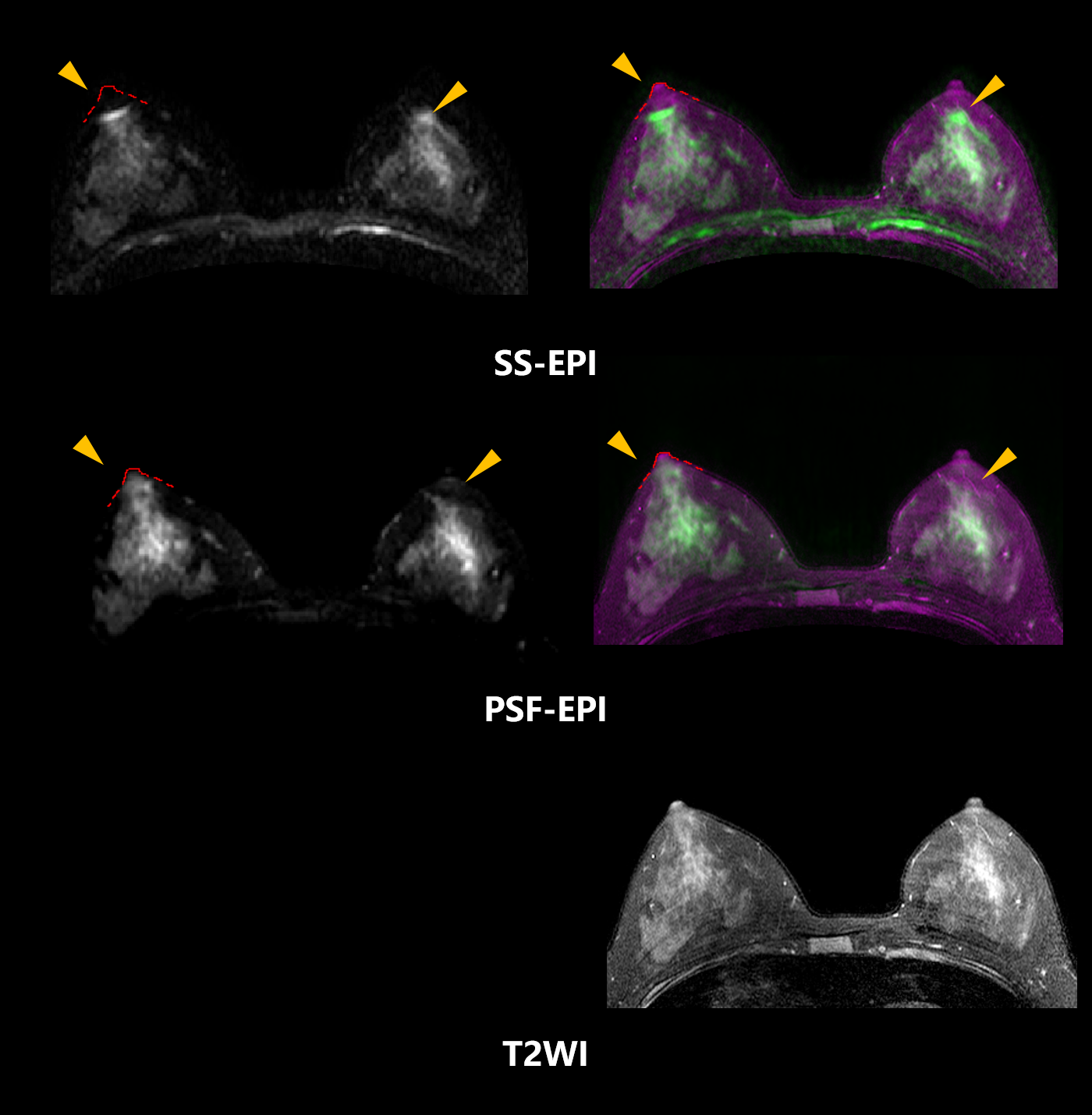
Figure 2 shows the b=0 s/mm2 images of a healthy female (22 y). In comparison with SS-EPI, there was better spatial consistency between PSF-EPI and the distortion-free T2WI. The serious geometric distortion around the nipple (arrow) on the b=0 s/mm2 image from SS-EPI was corrected using PSF-EPI.
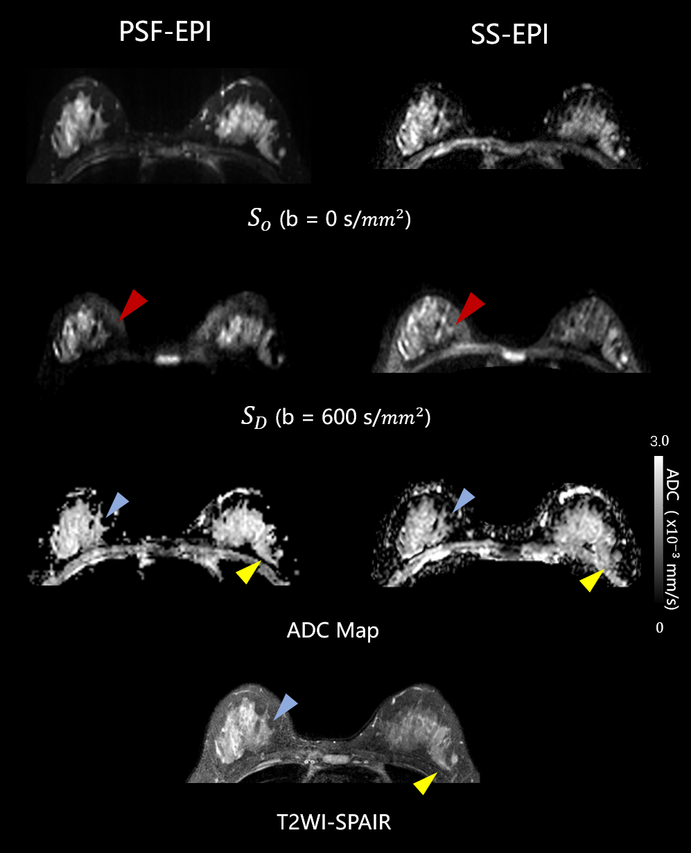
Figure 3 demonstrated that the DWI (b=600 s/mm2) image using PSF-EPI was not affected by the chemical shift artifact, which was, on the other hand, noticeable in the SS-EPI sequence due to poor fat suppression. It attributed to the good property of the PSF-EPI sequence that signals at different kpsf with the same ky position were acquired at the same echo time. ADC calculated from the PSF-EPI followed the anatomy more closely, and was slightly higher than that in SS-EPI.
Discussion
Breast DWI using PSF-EPI may be useful for breast tumor diagnosis since most tumors are prone to occur on the interface where distortion is severe due to field inhomogeneity.
Eddy current effects may cause spatial mismatch between images within a DWI sequence and result in false ADC values 1.The proposed PSF-EPI sequence has the superiority of little geometric distortion and chemical shift artifacts, thus avoiding such ADC inaccuracies. Slight motion-induced phase errors still occur despite of the correction, which is likely to be one of the reasons why PSF-DWI has a slightly higher ADC.
Furthermore, PSF-EPI DWI of the breast also has the great potential for lesion measurements since it can propagate ROIs directly from other MRI contrast, thus enhancing the quantitative accuracy of bilateral breast DWI exams.
Conclusion
In conclusion, breast DWI using PSF-EPI has the advantage of almost no distortion compared to that using SS-EPI, and could facilitate more accurate multi-parametric heterogeneity assessment. More patients will be recruited to evaluate this technique in the future.Acknowledgements
No acknowledgement found.References
1. Partridge S C, Mcdonald E S. Diffusion weighted MRI of the breast: Protocol optimization, guidelines for interpretation, and potential clinical applications. Magnetic Resonance Imaging Clinics of North America, 2013, 21(3):601.
2. Donz, Wang F, Reese TG, et al. Tilted-CAIPI for highly accelerated distortion-free EPI with point spread function (PSF) encoding. Magn Reson Med. 2018; 00:1-16.
3. Hancu I, Lee S, Hulsey K, et al. Distortion correction in diffusion-weighted imaging of the breast: Performance assessment of prospective, retrospective, and combined (prospective plus retrospective) approaches. Magnetic Resonance in Medicine, 2017,78(1):247-253.
4. Shotaro Kanao, Masako Kataoka, et al. High-resolution diffusion-weighted MRI of the breast using readout-segmented EPI and single-shot EPI. Imaging Med. 2017,9(6):185-190.
5. In M, Posnansky O, Speck O. High-resolution distortion-free diffusion imaging using hybrid spin-warp and echo-planar PSF-encoding approach. NEUROIMAGE, 2017,148:20-30.
6. Oh SH, Chung JY, In MH, Zaitsev M, Kim YB, Speck O, Cho ZH. Distortion correction in EPI at ultra-high-field MRI using PSF mapping with optimal combination of shift detection dimension. Magnetic resonance in medicine 2012;68(4):1239-1246.
7. In MH, Posnansky O, Beall EB, Lowe MJ, Speck O. Distortion correction in EPI using an extended PSF method with a reversed phase gradient approach. PloS one 2015;10(2):1-19.
8. In MH, Speck O. Highly accelerated PSF-mapping for EPI distortion correction with improved fidelity. Magnetic Resonance Materials in Physics, Biology and Medicine 2012;25(3):183-192.
9. Chung JY, In MH, Oh SH, Zaitsev M, Speck O, Cho ZH. An improved PSF mapping method for EPI distortion correction in human brain at ultra high field (7T). Magnetic Resonance Materials in Physics, Biology and Medicine 2011;24(3):179-190.
10.Robson MD, Gore JC, Constable RT. Measurement of the point spread function in MRI using constant time imaging. Magnetic resonance in medicine 1997;38(12):733-740.
11.Zaitsev M, Hennig J, Speck O. Point spread function mapping with parallel imaging techniques and high acceleration factors: Fast, robust, and flexible method for echo-planar imaging distortion correction. Magnetic resonance in medicine 2004;52(5):1156-1166.
12.In MH, Posnansky O, Speck O. PSF mapping-based correction of eddy-current-induced distortions in diffusion-weighted echo-planar imaging. Magnetic resonance in medicine 2016;75(5):2055-2063.
Figures