1872
A dual-slab 3D gradient echo spectroscopic imaging sequence with correction of respiration- and hardware-related frequency variations for bilateral evaluation of lipid composition in the breastPippa Storey1, Linda Moy1, and Sungheon G. Kim 1
1Radiology Department, New York University School of Medicine, New York, NY, United States
Synopsis
To enable rapid bilateral evaluation of lipid composition in the breast with high spatial resolution and easy integration into clinical workflow, we have developed a gradient echo spectroscopic imaging sequence, which consists of a simultaneous dual-slab 3D gradient echo imaging acquisition with 128 monopolar echoes. To correct for the frequency drift of the scanner over the course of the acquisition, as well as frequency fluctuations due to respiration, the sequence includes a quick frequency navigator every TR period. The navigator was found to improve spectral quality in all subjects.
Introduction
Recent studies have shown that a majority of breast cancer develops at the interface between fibroglandular tissue and adipose tissue. In particular, it has been found that postmenopausal women with invasive ductal carcinoma have a significantly higher percentage of saturated fat in their breast adipose tissue than postmenopausal women with only benign lesions [1]. To enable rapid bilateral evaluation of lipid composition in the breast with high spatial resolution and easy integration into clinical workflow, we have developed a gradient echo spectroscopic imaging sequence, which consists of a simultaneous dual-slab 3D gradient echo imaging acquisition with 128 monopolar echoes. No specialized shimming or voxel placement is required at the time of data acquisition, since spectra can be obtained for each voxel or region of interest within the 3D volumes after the exam by performing a Fourier transform along the echo dimension. To correct for the frequency drift of the scanner over the course of the acquisition, as well as frequency fluctuations due to respiration, the sequence includes a quick frequency navigator every TR period. The navigator consists of a short gradient echo train with 12 echoes and no phase- or partition-encoding, and a flip angle low enough to avoid altering the steady state.Methods
Data acquisition was performed in 8 women at 3T (TIM Trio, Siemens) using a 16-channel breast coil (Invivo). Imaging slabs with 2.8mm isotropic resolution and 18 slices per slab were prescribed over each breast as illustrated in Figure 1. Other parameters included: in-plane matrix size = 96x84, inter-echo spacing = 1.44ms, receiver bandwidth = 1410 Hz/pixel. A low FA of 10 degrees was chosen for the spectroscopic imaging acquisition to minimize T1 weighting. An even lower FA of 3 degrees was used for the frequency navigator to avoid disturbing the steady state. The total repetition time was 213ms, including 190ms per k-space line for the spectroscopic imaging acquisition and 23ms for the frequency navigator. The total acquisition time was 5:36. The phases of the k-space data from the spectroscopic imaging acquisition were corrected for frequency variation prior to reconstruction. A GRAPPA algorithm was used to unalias the data from the two breasts.Results
Figure 2 shows the frequency variation as a function of time over the course of the acquisition for a typical subject, as calculated from the frequency navigator. Note that it exhibits fluctuations due to respiration, superimposed on a gradual drift due to heating of the hardware. Figure 3 shows results from a central slice in the same subject with and without phase correction of the k-space data to compensate for the frequency variations. Note that the image with phase correction exhibits less artifact in the background, a longer FID in the tissue and narrower spectral peaks. The height of the principal spectral peak was used as a metric of spectral quality. The mean ratio of this height with and without phase correction was greater than one in each breast across all subjects. Averaged over both breasts, the mean ratio across all subjects was 1.093 +/- 0.036 (p = 7 x 10^-12)Discussion
We have developed a gradient echo spectroscopic imaging sequence to evaluate lipid composition bilaterally in the breast with high spatial resolution and easy integration into clinical workflow. The incorporation of a frequency navigator to compensate for respiratory-related frequency fluctuations and scanner-related frequency drift improved spectral quality in all subjects.Acknowledgements
This work was supported in part by NIH grants R01 CA219964 and P41 EB017183.References
1. Evaluation of Breast Lipid Composition in Patients with Benign Tissue and Cancer by Using Multiple Gradient-Echo MR Imaging. Radiology. 2016 Oct;281(1):43-53Figures
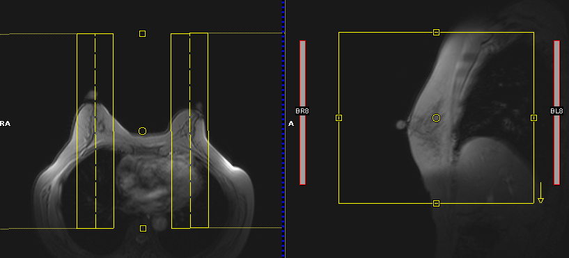
Figure 1: Graphic prescription of the simultaneous dual-slab gradient-echo spectroscopic imaging sequence in both breasts.
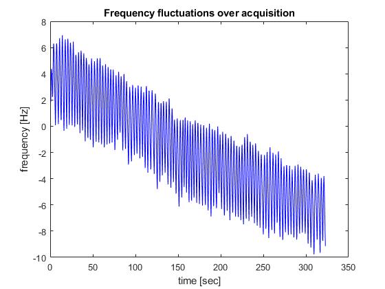
Figure 2: Frequency variations in one breast of the subject shown in Figure 1 over the course of data acquisition as calculated from the frequency navigator. Note the fluctuations due to respiration are superimposed on a drift due to the hardware.
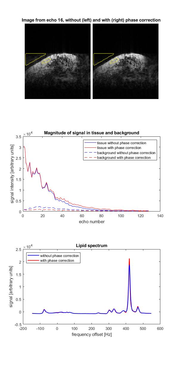
Figure 3: Results with and without phase correction in one breast of the subject shown in Figure 1, The top panel shows images from echo 16, and the second panel shows the signal magnitude as a function of echo number in the tissue and background ROIs. The lower panel shows the lipid spectrum from the tissue ROI.