1865
Diffusion kurtosis imaging and intravoxel incoherent motion analysis in comparison to a combination of multi-level diffusion coefficients in MR mammography of suspicious breast lesions1Medical Physics in Radiology, German Cancer Research Center (DKFZ), Heidelberg, Germany, 2Radiology, German Cancer Research Center (DKFZ), Heidelberg, Germany, 3Institute of Radiology, University Hospital Erlangen, Erlangen, Germany, 4Radiological Practice at the ATOS Clinic Heidelberg, Heidelberg, Germany, 5Radiology Center Mannheim (RZM), Mannheim, Germany
Synopsis
Diffusion-weighted imaging (DWI) is increasingly used in MR mammography for assessing malignancy of breast lesions. In particular, quantitative analysis of DWI data acquired with several b-values is gaining importance. Common data evaluation approaches include intravoxel incoherence motion (IVIM) and kurtosis imaging. The aim of this work was to evaluate the performance of kurtosis and IVIM based parameters in comparison to an approach combining several conventional diffusion coefficients measured with different b-values. The dataset comprised 198 examinations of patients with suspicious mammography findings. The simple approach using several ADCs showed a similar performance as the combination of kurtosis and IVIM.
INTRODUCTION
Diffusion-weighted imaging (DWI) provides a high performance in discriminating between malignant and benign breast lesions. The apparent diffusion coefficient (ADC) is a robust parameter, which allows quantification of diffusion restrictions1. Diffusion kurtosis imaging can provide additional information about the heterogeneity of the tissue2. Moreover, the intravoxel incoherent motion (IVIM) model can yield parameters quantifying blood flow in capillaries3. This work addresses the question of whether using these specific models is superior in discriminating benign from malignant breast lesions compared to combining several ADC values calculated at different b-value levels from a multi b-value acquisition.METHODS
Prospectively acquired DWI datasets of patients with suspicious BI-RADS 4/5 lesions revealed during X-ray mammography screening and indication for breast biopsy were involved in this IRB approved study. Histology was used as ground truth for assessing malignancy. In total, 198 patients were examined with 1.5T MRI scanners at two study centres prior to biopsy. In Group A (105 patients), a single-shot EPI sequence at a Philips Ingenia MRI scanner was used (acq. time 3:41 min), whereas in Group B (93 patients), segmented EPI at Siemens Aera MRI-scanner was applied (3 readout segments, acq. time 6:44 min). Multiple b-values (0, 100, 750 and 1500 s/mm2) were used in both DWI sequences. The voxel size was 2.5 x 2.5 x 3 mm³. Regions of interest were delineated on the image with the highest b-value where the lesion was visible while taking the T2 images visually into account. The first predicting model, based on logistic regression, was constructed from 2 parameters: the apparent diffusion coefficient (Dapp), the apparent kurtosis coefficient (Kapp) obtained from the diffusion kurtosis equation $$S(b)=S_0\cdot e^{\left(-b D_{app}+\frac{1}{6}b^2{D_{app}}^2K_{app}\right)}$$ using 3 b-values (100,750 and 1500 s/mm2). The second approach, again using logistic regression, combined the kurtosis-based parameters Dapp and Kapp with the perfusion fraction f calculated by applying the IVIM equation $$S(b)=S_0\left[(1-f)\cdot e^{-bD}+f\cdot e^{-b\left(D^*+D\right)}\right].$$ Due to the instability of the fitting, the f was calculated in two steps as $$f=\frac{S_0-{S_0}'}{S_0}$$ where S0' was estimated from $$S(b)=S_0'\cdot e^{-b\cdot ADC_{100-750}}$$ and ADC100-750 was obtained employing the b-values = 100 and 750 s/mm2. For the third approach, three individual ADC values were calculated using $${ADC}_i=-\frac{1}{b_i}\ln\left( \frac{S(b_i)}{S(b=0)}\right)$$ for b1=100 s/mm2, b2=750 s/mm2, b3=1500 s/mm2 thus combining the signal without diffusion weighting and that acquired with b-values larger than zero. The resulting three ADC-values were then evaluated using logistic regression.
RESULTS
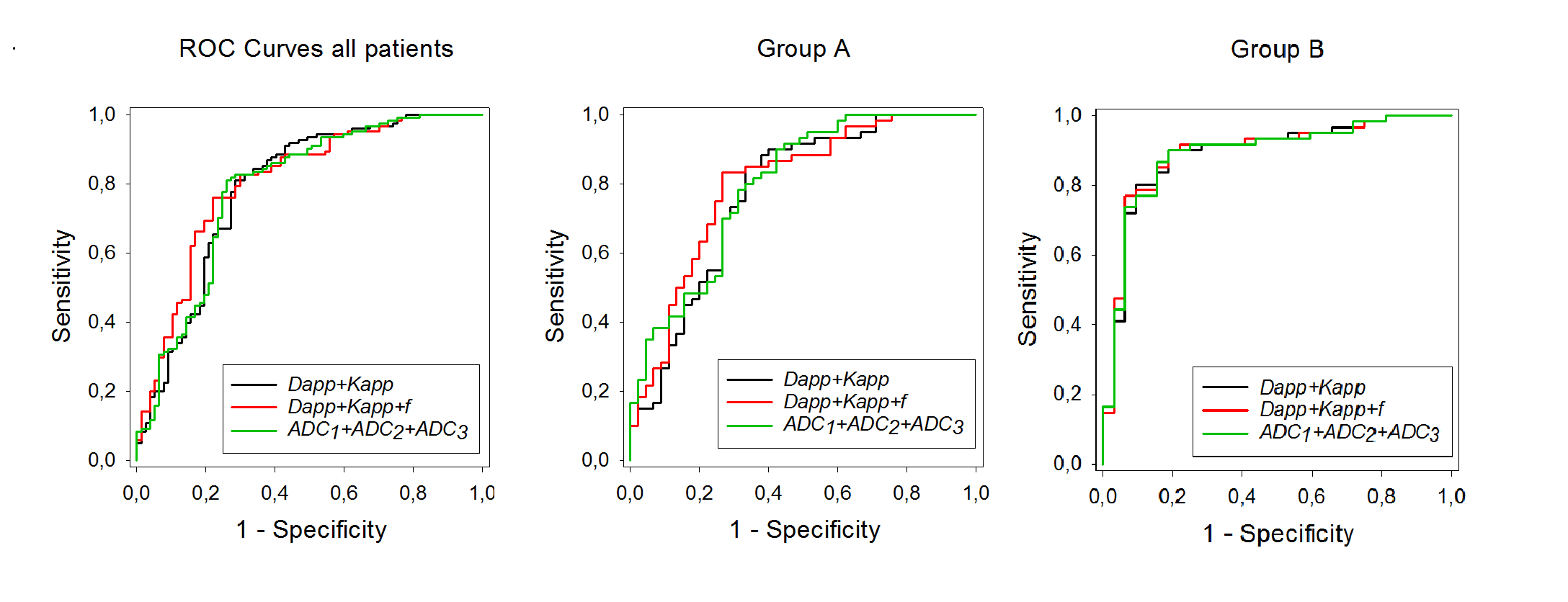
The predicting ability of the models based on the logistic regression was assessed by the analysis of the receiver operating characteristic (ROC) curve shown in Figure 1. The first approach using Dapp and Kapp yielded an area under the curve (AUC) of 0.79 (95%CI 0.72-0.86), the combination of Dapp, Kapp and f resulted in an AUC value of 0.81 (95%CI 0.74-0.87), while the evaluation of the three ADC-values yielded the AUC 0.79 (95%CI 0.72-0.86). Thus, no significant difference in the performance of the different evaluation approaches could be observed (p>0.18). The AUC when analyzing f alone was only 0.505 (95%CI 0.419-0.590).
For the individual analysis of the data obtained in the two study centers, for Group A and the three-parameter combination (Dapp, Kapp, f), the AUC 0.80 (95%CI 0.71-0.89) was obtained, while the AUC 0.80 (95%CI 0.71-0.88) was calculated when using three individual ADCs. For Group B, the respective AUC values were: 0.89 (95%CI 0.82-0.97) and 0.89 (95%CI 0.82-0.96). Performing the calculation using Dapp and Kapp only resulted in AUC 0.77 (95%CI 0.68-0.87) for Group A and AUC 0.89 (95%CI 0.82-0.97) in Group B. No significant difference in the performance could be observed in the individual groups, as well.
DISCUSSION and CONCLUSION
This work addressed the question whether the kurtosis and IVIM parameters contain inherent properties when analyzing the signal in suspicious breast lesion that may be superior to a simplified approach analyzing conventional ADC values with different b-values using logistic regression. In this analysis, no advantage in diagnostic accuracy of the more advanced models could be observed and simply using a combination of different ADC values seemed to be a viable approach. For our dataset, the performance of f was particularly bad. It has to be noted however, that an abbreviated protocol was used with the aim of short measurement time for integration into clinical routine, which is far from being optimal for IVIM and kurtosis analysis regarding SNR and employed b-values. Furthermore, it remains an open question whether using the available measurement time to acquire only two b-values thus resulting in only one ADC value would lead to similar results as the methods presented here.Acknowledgements
No acknowledgement found.References
1. Bickelhaupt S, Steudle F, Paech D, Mlynarska A, Kuder TA, Lederer W, Daniel H, Freitag M, Delorme S, Schlemmer HP, Laun FB. On a fractional order calculus model in diffusion weighted breast imaging to differentiate between malignant and benign breast lesions detected on X-ray screening mammography. PLoS One. 2017 Apr 28;12(4):e0176077.
2. Jensen JH, Helpern JA, Ramani A, Lu H, Kaczynski K., Diffusional kurtosis imaging: the quantification of non-gaussian water diffusion by means of magnetic resonance imaging., Magn Reson Med. 2005 Jun;53(6):1432-40.
3. Le Bihan D, Breton E, Lallemand D, Aubin ML, Vignaud J, Laval-Jeantet M. Separation of diffusion and perfusion in intravoxel incoherent motion MR imaging. Radiology. 1988 Aug;168(2):497-505.
Figures