1864
Intravoxel incoherent motion imaging in the prediction of aggressiveness of T1 breast carcinoma patients1Radiology, The First affiliated hospital of Dalian Medical University, Dalian, China, 2GE Healthcare, China, Beijing, China
Synopsis
Intravoxel incoherent motion (IVIM) imaging provides quantitative measurement of ADCslow for cellularity and ADCfast and ffast for vascularity. It is helpful for the differentiation between benign and malignant breast lesions. This study concerned perfusion as well as diffusion parameters of T1 breast carcinoma lesions using IVIM imaging based on the biexponential analysis and then compared these parameters from T1-weighted, T2-weighted and contrast-enhanced MR images on the classification of high and low aggressiveness of T1 breast carcinomas.
Synopsis
Intravoxel incoherent motion (IVIM) imaging provides quantitative measurement of ADCslow for cellularity and ADCfast and ffast for vascularity. It is helpful for the differentiation between benign and malignant breast lesions. This study concerned perfusion as well as diffusion parameters of T1 breast carcinoma lesions using IVIM imaging based on the biexponential analysis and then compared these parameters from T1-weighted, T2-weighted and contrast-enhanced MR images on the classification of high and low aggressiveness of T1 breast carcinomas.Purpose
To identify IVIM features in the prediction of tumor aggressiveness of T1 breast carcinoma patients.Methods
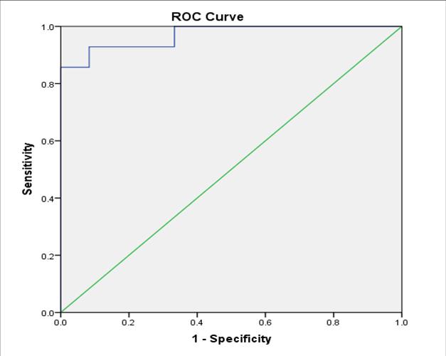
In this prospective study, a total of 56 female patients with 57 T1 breast carcinomas were enrolled and informed consent was acquired from each patient. Specifically, group 1 included 29 patients with low aggressive pathological subtype and group 2 included 28 patients with high aggressive pathological subtype. Tumor aggressiveness was defined according to the surgical histopathology. IVIM (18 b value), T1-weighted, T2-weighted and contrast-enhanced MR sequences were acquired. Tumor size, DWI parameters (ADCstandard, ADCslow, ADCfast and ffast) and MRI features of lesions were obtained. The parameters were compared between two groups and the diagnostic performance was quantified with ROC curve. A multivariate logistic regression model was developed to identify features that were independently predictive for tumor aggressiveness.Results
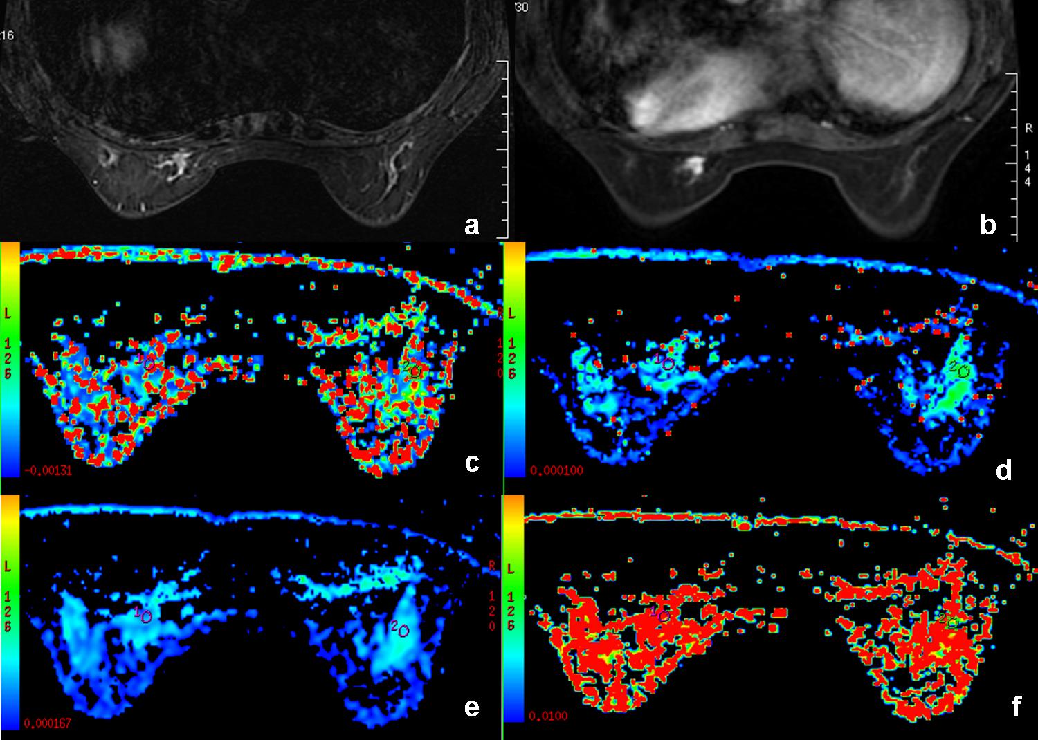
There was no significant difference in age and tumor size between two groups (p=0.257, 0.36). The lesions of high aggressive pathological subtype can be significantly differentiated from the lesions of low aggressive pathological subtype from ADCslow (P=0.008), enhancement shape (P<0.001), tumor margin on delayed contrast-enhanced images (P<0.001), and internal enhancement (P=0.018) (Fig 1). The highest area under the curve (AUC) was acquired by combining ADCslow and tumor margin on contrast-enhanced images with an optimal threshold of 0.276×10⁻³mm2 (Fig 2).Acknowledgements
This work was supported by a grant of the Basic Scientific Research Projects of the Universities in Liaoning Province (LQ2017013).References
[1] Chen W, Zheng R, Baade PD, et al. Cancer statistics in China, 2015. CA Cancer J Clin. 2016, 66(2): 115-132.
[2] Lee YJ, Kim SH, Kang BJ, et al. Intravoxel incoherent motion (IVIM)-derived parameters in diffusion weighted MRI: Associations with prognostic factors in invasive ductal carcinoma. J Magn Reson Imaging. 2017, 45(5): 1394-1406.
[3] Colleen Bailey, Bernard Siow, Eleftheria Panagiotaki, et al. Microstructural models for diffusion MRI in breast cancer and surrounding stroma: an exvivo study. NMR Biomed. 2017, 30(2): e3679.
[4] Kim Y J, Ko K, Kim D H, et al. Intravoxel incoherent motion diffusion-weighted MR imaging of breast cancer: Association with histopathological features and subtypes. Br J Radiol. 2016, 89(1063): 20160140.
Figures