1861
Effect of DWI sequence on geometric measurement errors in a breast phantom1University of California San Francisco, San Francisco, CA, United States, 2National Institute of Standards and Technology, Boulder, CO, United States, 3University of California Los Angeles, Los Angeles, CA, United States
Synopsis
Previously, when using the UCSF/NIST breast phantom, we discovered an x-direction distortion in DW-EPI and the resulting ADC maps that was dependent on spatial location within the scanner and was also present in patient images. The distortion was unaffected by switching the phase encode direction and did not change with increasing b-value, and thus was most likely not due to gradient nonlinearity nor eddy currents. Here, we demonstrate that the distortions decrease when using multi-shot EPI, which is less sensitive to off-resonance distortion, and change direction when reversing the gradient polarity.
Introduction
Previously, when using the UCSF/NIST breast phantom, we discovered a spatial distortion in diffusion-weighted echo planar imaging (DW-EPI) and the resulting apparent diffusion coefficient (ADC) maps that was dependent on spatial location within the scanner (patient left or right) and was also present in patient images1. The distortion was an x-direction stretching on one coil side and compression on the other coil side. The distortion was unaffected by switching the phase encode direction and did not change with increasing b-value. Thus, the stretching/compression distortion was most likely not due to gradient nonlinearity nor eddy currents. One hypothesized cause of the distortion was: B0 inhomogeneity effects due to the magnet, coil or shim settings2.
Now there are additional image correction techniques available for breast imaging that may be utilized to investigate potential causes of the stretching/compression distortion, including multi-shot EPI techniques3,4 and gradient polarity reversal5. For this study, we imaged one UCSF/NIST breast phantom on two different vendor 3T systems using both single-shot and multi-shot EPI acquisitions. If the distortion is due to B0 inhomogeneity, it would be expected to decrease when using multi-shot EPI techniques, which are less sensitive to off-resonance distortion.
Methods
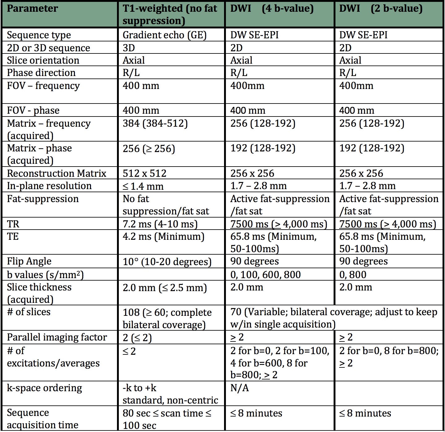
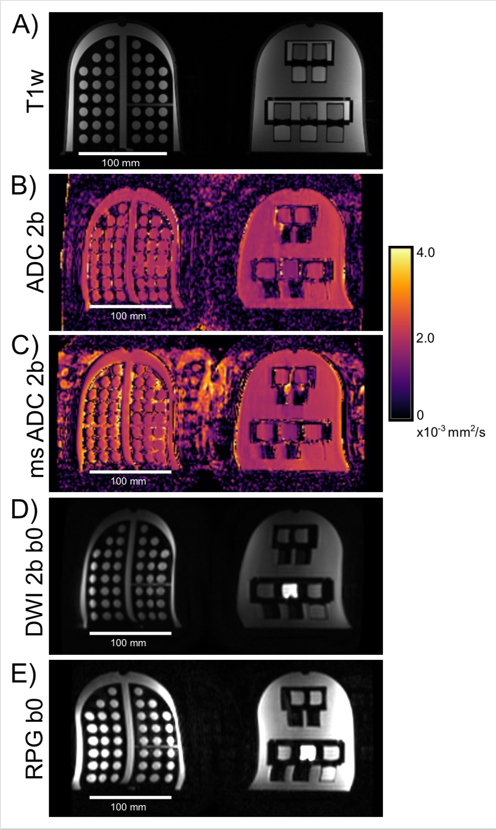
Using the commercial version of the UCSF/NIST breast phantom (QalibreMD), we collected T1-weighted, DWI 2 b-value, DWI 4 b-value, DWI 2 b-value multi-shot and B0 map image sets (Fig. 1) on Site 1: Siemens 3T Skyra with a 16-channel breast coil (Siemens) and Site 2: GE Medical Systems 3T Signa Premier with a 16-channel breast coil (Invivo). To acquire test-retest images, the phantom was removed from the breast coil and then repositioned at least 20 minutes later on the same day. The diffusion phantom unit was placed on the patient right and the T1 phantom unit was placed on the patient left. Axial MRI data were acquired in the standard breast imaging orientation: in feet first prone. During one scan session on the GE 3T system, reverse polarity gradient (RPG) images were also acquired.
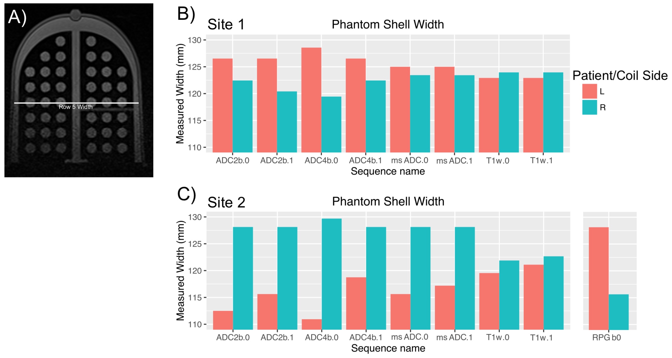
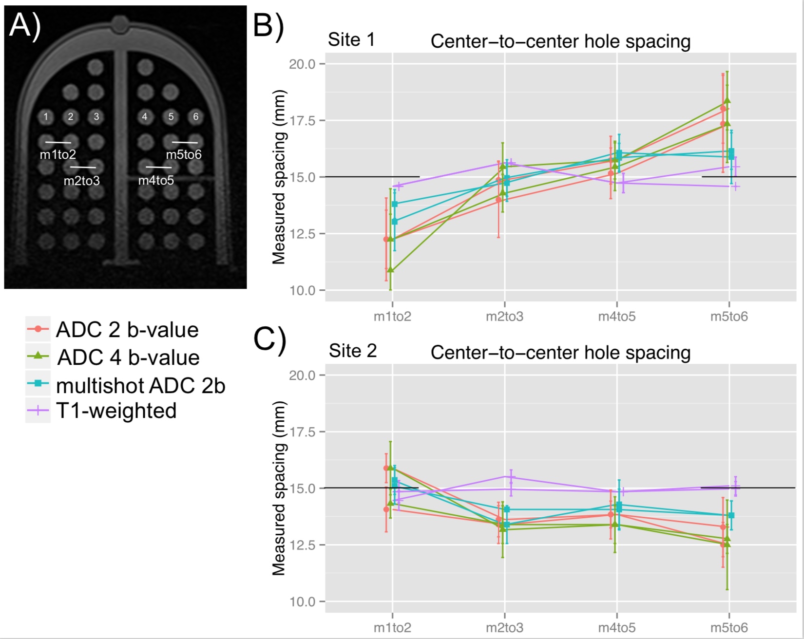
Distortion measurements were made using an automatic in-house processing algorithm written in Python and confirmed manually. First, the interior width of the phantom was measured on both the diffusion and T1 units at the location of the fifth row of the geometric distortion plate (Fig. 2). Second, the center-to-center spacing was measured across rows of the geometric distortion array (Fig. 3). The average and standard deviation of these measurements across the seven rows with multiple holes are reported. From the engineering drawings, the fabricated center-to-center spacing is 15 mm.
Results
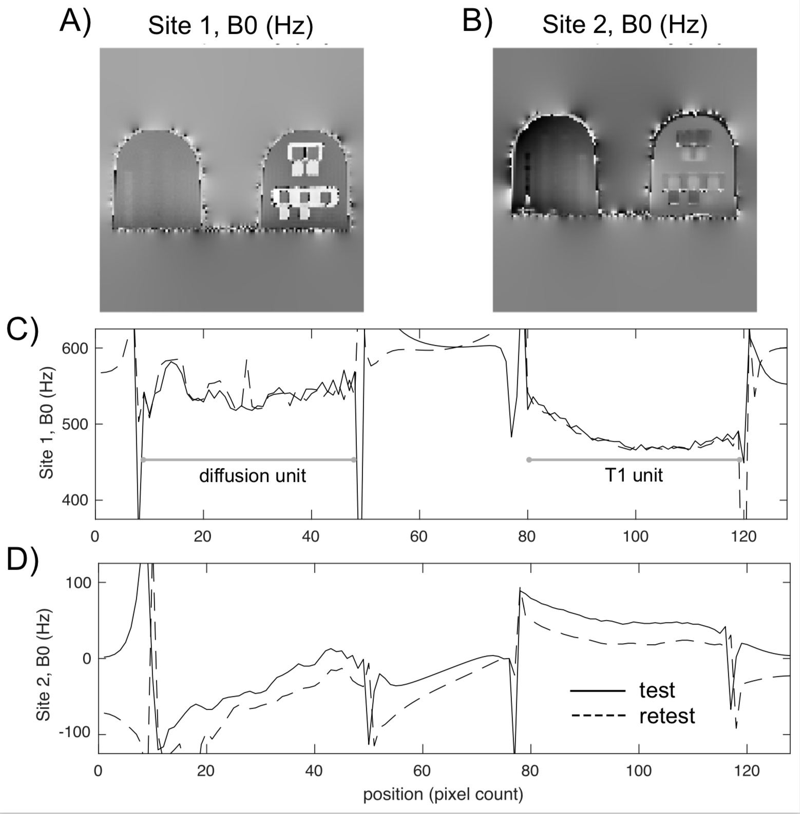
Data collected in the commercialized UCSF/NIST breast phantom demonstrated a stretching/compression distortion in the x-direction dependent on R/L spatial location within the magnet (Fig. 2). In addition, the magnitude of the distortion decreased when using multi-shot EPI techniques and switched direction when using RPG (Fig. 3). The B0 magnitude varied depending on system and spatial location within the magnet (Fig. 4). Finally, examining just the diffusion phantom unit, the center-to-center hole spacing was not linear across the geometric distortion grid at Site 1 (Fig. 5). At both sites, the T1-weighted data was closest to the fabricated 15 mm spacing, the single-shot EPI ADC maps were most distorted, and the multi-shot EPI images were only moderately distorted.Discussion
We demonstrated a potential B0 dependence of the stretching/compression distortion, because the magnitude of the distortion decreased when using multi-shot EPI sequences, which are less susceptible to B0 inhomogeneity. Also, at Site 2, we were able to reverse the stretching/compression distortion direction by reversing the gradient polarity. Finally, we observed in the B0 maps that the coil side with the greater B0 value was the side with the stretch distortion.
We were not able to attribute a source of the B0 inhomogeneity; possible causes are the main magnet, shim settings, the breast coil, or other objects within the field. Also, the width measurement for each unit of the phantom was of the interior of a soft shell. There could be measurement variation due to the soft sides of the shell, and this is one possible cause of the measurement variation in the T1-weighted images. We are working with other breast imaging sites to collect additional data.
Conclusion
Geometric distortions hinder the co-registration of EPI images to images from other clinically-relevant sequences, such as T1-weighted DCE, which is useful for multi-parametric breast tumor characterization6. It is important to be aware of geometric distortion artifacts, and the UCSF/NIST breast phantom is a useful tool to identify and characterize these artifacts and test techniques to reduce their effects.Acknowledgements
This work was supported in part by NIH R01 CA132870 and NIH U01 CA225427. The authors appreciate thoughtful conversations with Scott Hinks (GE Medical Systems).References
[1] Keenan KE, Peskin AP, Wilmes LJ et al. Variability and bias assessment in breast ADC measurement across multiple systems. JMRI. 2016; 44(4):846-855.
[2] Jezzard P, Balaban RS. Correction for geometric distortion in echo planar images from B0 field variations. MRM. 1995; 34(1):65-73.
[3] Porter DA, Heidemann RM. High resolution diffusion-weighted imaging using readout-segmented echo-planar imaging, parallel imaging and a two-dimensional navigator-based reacquisition. MRM. 2009; 62(2):468-475.
[4] Chen NK, Guidon A, Chang HC, Song AW. A robust multi-shot scan strategy for high-resolution diffusion weighted MRI enabled by multiplexed sensitivity-encoding (MUSE). NeuroImage. 2013; 72:41–47.
[5] Holland D, Kuperman JM, Dale AM. Efficient correction of inhomogeneous static magnetic field-induced distortion in Echo Planar Imaging. Neuroimage. 2010; 50(1):175-183.
[6] Partridge SC, Zhang Z, Newitt DC, et al. Diffusion-weighted MRI findings predict pathologic response in neoadjuvant treatment of breast cancer: the ACRIN 6698 multicenter trial. Radiology. 2018. https://doi.org/10.1148/radiol.2018180273
Figures