1838
Advanced diffusion-weighted imaging with multiple parameters in differential diagnosis of prostate transitional zone carcinoma and benign prostatic hyperplasia1Baoji Central Hospital, Baoji, China, 2GE Healthcare China, Beijing, China
Synopsis
Conventional MRI sequences and DWI sequences are difficult to identify prostate transitional cell carcinoma and benign prostatic hyperplasia. In this study, traditional DWI sequences, intravoxel incoherent motion diffusion weighted imaging (IVIM-DWI), stretch index DWI sequences and DKI sequences were used to compare the accuracy of these sequence parameters in the differential diagnosis of prostate cancer and benign prostatic hyperplasia. The results showed that ADC-slow, DDC, Md, Da, Dr, MK, Ka and Kr were helpful in differentiating prostate cancer from benign prostatic hyperplasia, and MK value had the highest accuracy in differentiating prostate cancer from benign prostatic hyperplasia.
Introduction
Prostate cancer is one of the most common malignant tumors among men. DWI sequences have been widely used in the diagnosis and differential diagnosis of prostate cancer, and prostate Imaging-Reporting and data system (PI-RADS) has been made avaibale1. Transitional zone is the most common site for benign prostatic hyperplasia (BPH). It is difficult to differentiate transitional zone cancer from hyperplastic nodules on conventional MRI and DWI images. Conventional diffusion weighted imaging is based on mono-exponential model and hence may be restricted in the diffusion information. In this work, advanced diffusion models including intravoxel incoherent motion (IVIM-DWI), stretched exponential DWI and Diffusion Kurtosis Imaging (DKI) in the differential diagnosis of prostate transitional zone carcinoma and prostatic hyperplasia, as compared to conventional DWI.Materials
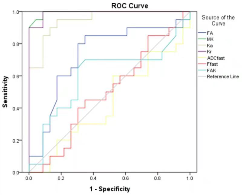
This retrospective study was approved by local ethical committee and consent forms were waived. 35 cases of prostate transitional carcinoma and 40 cases of benign prostatic hyperplasia were enrolled. All participants underwent MR exams on a whole body 3.0T scanner (Discovery MR750, GE, WI). MRI routine sequence, mono-exponential DWI, IVIM DWI(Sequence parameters are shown in Table 1), stretched exponential DWI and DKI scan were performed. Detailed scan parameters can be found in Table 1. The apparent diffusion coefficient (ADC) from mono-exponential DWI; slow diffusion ADC value (ADC-slow), rapid diffusion ADC value (ADC-fast), perfusion fraction (F-fast) from IVIM-DWI; water molecular diffusion heterogeneity index (Alpha), distributed diffusion coefficient (DDC) from stretched exponential DWI, diffusion anisotropy (FA), mean diffusion coefficient (MD), axial diffusion coefficient (Da), radial diffusion coefficient (Dr), diffusion kurtosis anisotropy (FAK), average diffusion kurtosis (MK), axial diffusion kurtosis (KA) and radial diffusion kurtosis (Kr) from DKI were analyzed. Measured diffusion markers were analyzed by T test, and ROC curve was obtained. The diagnostic efficiency of each parameter was investigated, and the diagnostic threshold was determined.Results
Lower values of ADC, ADC-slow, DDC, alpha, Md, Da and Dr were observed in the cancer group than those in the hyperplastic group (p<0.01). These areas under the curve(AUC) were 0.912, 0.985, 0.991, 0.796, 1.445, 0.963 and 0.984 respectively. The diagnostic thresholds were 0.836×10⁻³mm2/s, 0.785×10⁻³mm2/s, 0.882×10⁻³mm2/s, 0.705, 1.36um2/ms, 1.65um2/ms and 1.16um2/ms respectively. The FA, MK, Ka and Kr values in the cancer group were higher than those in the proliferative group (p<0.01), These AUC were 0.730, 0.995, 0.948 and 0.991 respectively, and the diagnostic thresholds were 0.193, 0.835, 0.824, 0.793 respectively. The diagnostic efficiency of ADC-slow, DDC, Md, Da, Dr, MK, Ka, Kr was high, and the sensitivity was 100%, 95%, 95%, 100%, 90%, 100%, 95%, 100% respectively. The specificity was 91.3%, 91.3%, 95.7%,91.3%,82.6%, 91.3%, The accuracy of ADC-fast, F-fast and FAK is relatively low. (The results are shown in Figure1, Table2, Figure2 and Table3. )Discussion
In addition to the mono-exponential model based perfusion information reflected in DWI. IVIM biexponential model can simultaneously reflect the diffusion and perfusion of water molecules in tissues. ADC-slow eliminates the effect of blood perfusion and can more truly reflect the diffusion of water molecules in tissues 2. Tensile index model can reflect the heterogeneity of water molecular diffusion and the average diffusion rate in voxels. DKI is based on a non-Gaussian diffusion model, which may better reflect the complex microstructure 3. The above model has been applied in the diagnosis and differential diagnosis of various tumors 4,5, better diagnostic performance was observed with the above model in differentiating prostate transitional zone cancer and hyperplasia. In this study, we found that the AUC of ADC-slow, DDC, Md, Da, Dr, MK, Ka, Kr was larger than that of ADC,FA and Alpha, And the AUC of MK was the largest.Conclusion
Advanced diffusion models including IVIM, DKI and tensile index showed better efficacy in differential diagnosis of prostate transitional zone carcinoma from benign prostatic hyperplasia, compared to conventional diffusion weighted imaging.Acknowledgements
No acknowledgement found.References
1.Weinreb JC, Barentsz JO, Choyke PL, et al. PI-RADS prostate imaging-reporting and data system: 2015, version 2. Eur Urol, 2016, 69(1): 16-40.
2.LE BIHAN D, BRETON E, LALLEMAND D, et al. MR imaging of intravoxel incoherent motions: application to diffusion and perfusion in neurologic disorders[J]. Radiology, 1986, 161(2): 401-407.
3.Jensen J H, Helpern J A. MRI quantification of non-Gaussian water diffusion by kurtosis analysis[J].NMR BIOMED,2010, 23(7):698-710.
4.Jiang R,Jiang J,Zhao L,et al. Diffusion kurtosis imaging can efficiently assess the glioma grade and cellular proliferation[J].Oncotarget, 2015,6(39):42380-42393.
5.Iima M,Le BD.Clinical intravoxel incoherent motion and diffusion MR imaging:Past,present,and future[J].Radiology,2016,278(1):13-32.
Figures