1834
Comparison of detection rate of prostate cancer between mpMRI guided in-bore biopsy and routine TRUS biopsy1Department of NMR and MRI Facility, All India Institute of Medical Sciences, New Delhi, India, 2Department of Radio-diagnosis, All India Institute of Medical Sciences, New Delhi, India, 3Department of Radio-diagnosis, IRCH, All India Institute of Medical Sciences, New Delhi, India, 4Department of Urology, All India Institute of Medical Sciences, New Delhi, India, 5Department of Pathology, All India Institute of Medical Sciences, New Delhi, India
Synopsis
The present study demonstrated the role of mpMRI guided in-bore biopsy for detection of prostate cancer (PCa). Patients were recruited based on PSA > 4 ng/ml and abnormal DRE. The PCa detection rate between the two groups of patients, namely 25 patients (Group I) who underwent in-bore biopsy and 73 patients (Group II) who underwent TRUS biopsy were compared. The PCa detection rate of in-bore targeted biopsy was 52% compared to 34.3% for TRUS biopsy. Our data also indicated that in-bore MR targeted biopsy significantly increased the detection rate of PCa when compared with the standard 12 core TRUS biopsy.
Purpose: To evaluate the role of mpMRI guided in-bore biopsy for detection of prostate cancer and compare the detection rate of prostate cancer using in-bore and TRUS biopsy.
Introduction: Prostate cancer (PCa) is the second most common cancer among men and more than 1.1 million new cases are reported worldwide every year1. PCa screening is performed with a digital rectal examination (DRE) and the serum prostate specific antigen (PSA) level. The aim of this study is to carry out mpMRI for finding suspicious areas of malignancy within the prostate and target these suspicious lesions based on PIRADS scores 4 or 5. Further, the detection rate of PCa is compared using in-bore and TRUS biopsy.
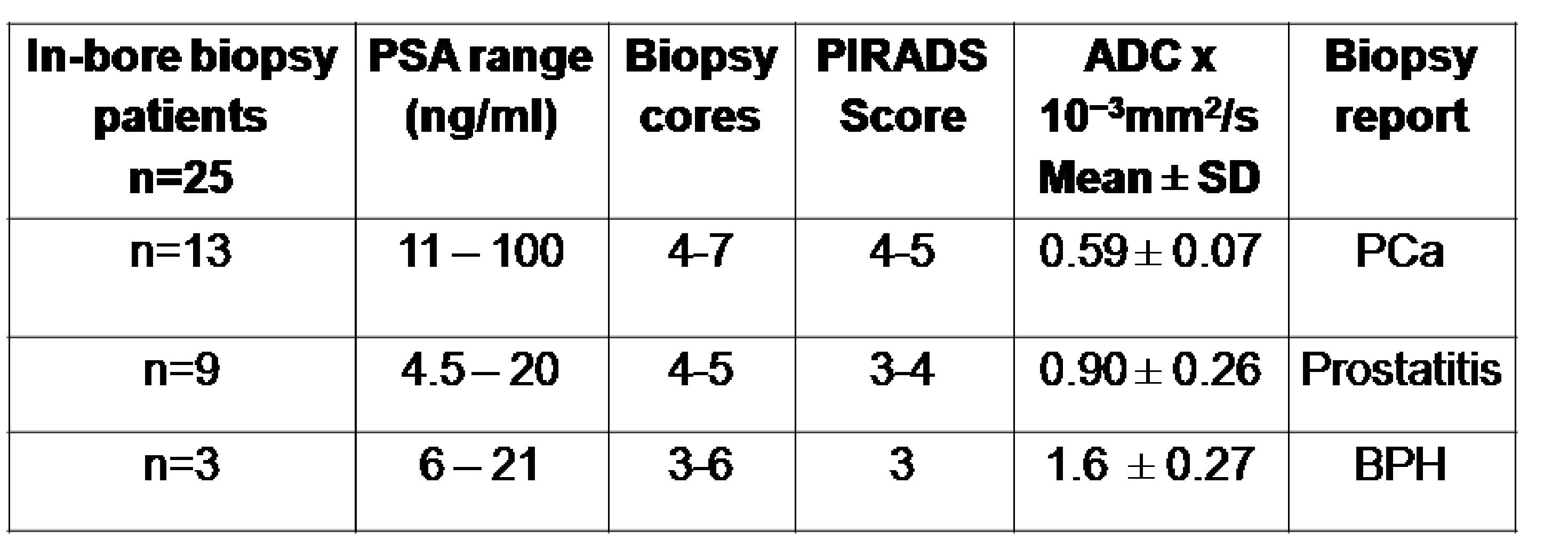
Methods: Patients were recruited based on PSA > 4 ng/ml and abnormal DRE. MR investigations were carried out at 3.0 T MRI system (Achieva, Philips, Netherlands), using mp-MRI (T1-WI, T2-WI, DWI and DCE-MRI). Patients were divided in to two groups: Group I consists of 25 patients, (age: 52 – 70 years, PSA range 4.5 to 100 ng/ml) who underwent in-bore biopsy while Group II had 73 patients who underwent only TRUS biopsy procedure. All patients had mpMRI Protocol. T2W images were acquired with the following parameters: repetition time (TR) of 6100 ms, echo time (TE) of 100 ms, field of view = 240 x 240 mm and slice thickness 3 mm, Further, all had diffusion weighted images acquired using seven b-values 0, 200, 400, 800, 1000, 1500 and 2000 s/mm2. Apparent diffusion coefficient (ADC) of tumor regions was calculated from ADC map. For DCE-MRI, 20 ml Gadolinium was injected at a rate of 3ml/s and T1W images were obtained every 2-5 s through the prostate. Group I patients underwent in-bore biopsy on the suspicious areas directly with in the bore of the magnet using a DynaTrim (Invivo, USA) biopsy device with needle guide and DynaCad software. Biopsy was carried out by transrectal approach and localization of lesion was confirmed using MR imaging before targeted biopsy. Three to six cores were collected from the suspicious areas. All lesions were assigned a score based on PI-RADS classification system. For Group II patients only routine 12 core TRUS biopsy was carried out by transrectal approach. Institute ethics committee approved the study and informed consent was taken from all subjects.
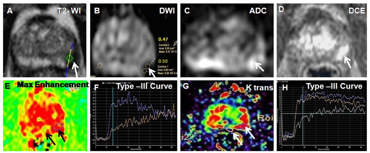
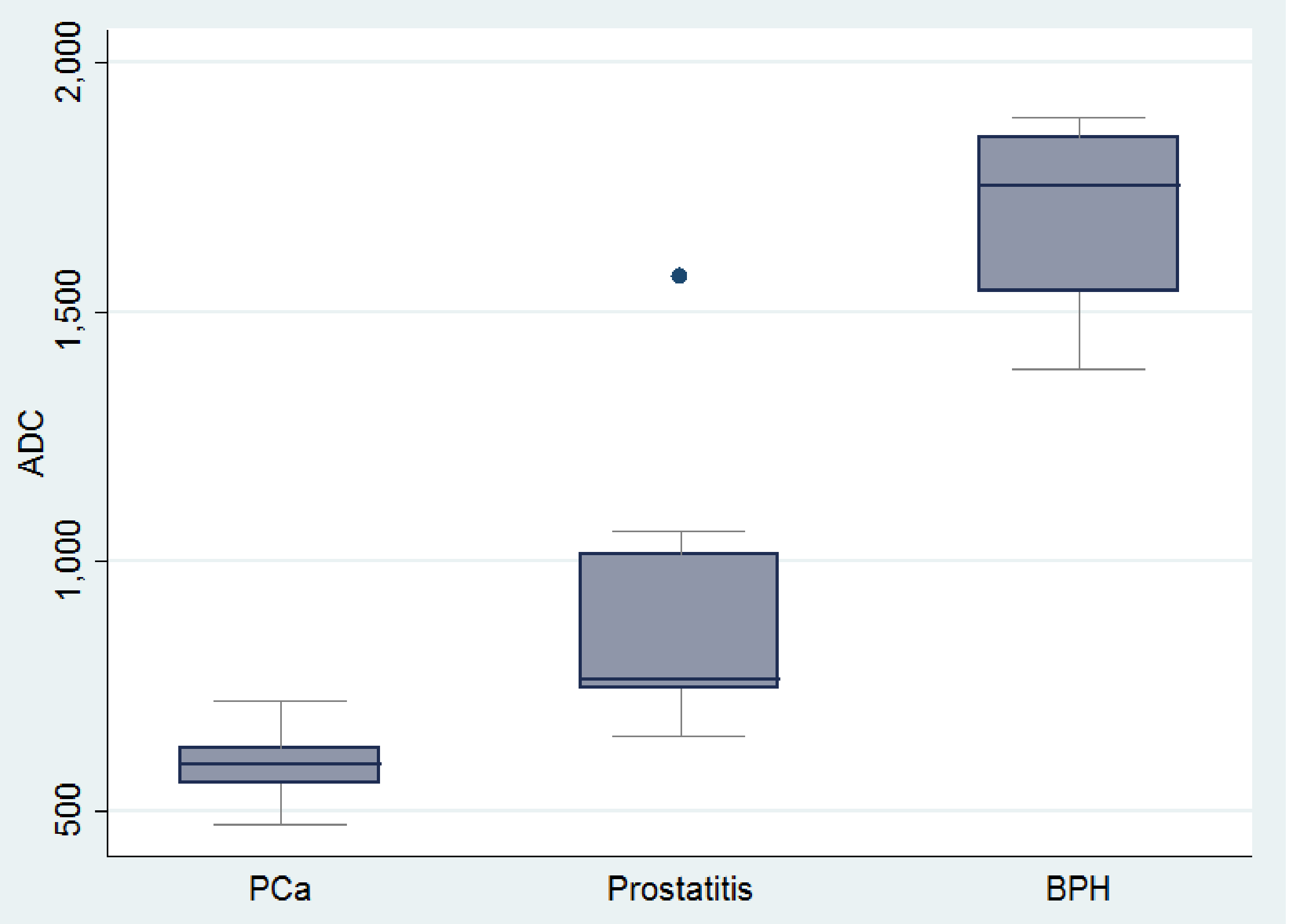
Results: Our study demonstrated that mpMRI accurately identified suspicious areas of lesion and MR guided in-bore biopsy helped to target these areas with accuracy (Figure 1). These targeted lesions with PIRADS scores (4 or 5) were correlated with the histopathological findings. The box plot of mean ADC showed significant difference between PCa, benign prostatic hyperplasia (BPH) and prostatitis (Figure 2). The mean ADC for malignant lesions (0.59 ± 0.07 x 10⁻³ mm2/s) was lower compared to prostatitis (0.90 ± 0.26 x 10⁻³mm2/s,) and BPH (1.6 ± 0.27 x 10⁻³mm2/s). The use of ADC may reduce the number of false positive findings as well as unnecessary biopsies cores.The PCa detection rate between the two groups of patients, namely 25 patients (Group I) who underwent in-bore biopsy and 73 patients (Group II) who underwent TRUS biopsy were compared. The PCa detection rate of in-bore targeted biopsy was 52% compared to 34.3% for TRUS biopsy. Histopathological findings of 25 patients who underwent MRI guided biopsy revealed that 13 were positive for cancer with a high Gleason score 7(4+3) and 8(4+4), while Nine patients had prostatitis and the rest 3 patients had BPH (Table1). In 73 patients who underwent TRUS biopsy, 25 were found to be positive for PCa. The rest 48 patients had prostatitis and BPH. Prostatitis and BPH have altered cellular density and interstitial space of tissues.
Discussion: In this study we demonstrated that in-bore biopsy of targeting suspicious areas of lesions shown by mpMRI improves the accuracy and increased the detection of PCa in men with elevated PSA and abnormal DRE. Our data also indicated that DWI may reduce the number of false positive findings and it also helps to reduce unnecessary biopsies. In-bore MR targeted biopsy significantly increased the detection rate of prostate cancer when compared with the standard 12 core TRUS biopsy.
Conclusion: mpMRI guided in-bore biopsy has the potential to reduce unnecessary biopsy cores and increase the detection of PCa.
Acknowledgements
NRJ thanks SERB, Government of India funding under FIST program (SR/FST/LSI-569/2013) and for J.C. Bose Fellowship.References
(1). Ferlay J, et al. Cancer incidence and mortality worldwide: sources, methods and major patterns in GLOBOCAN 2012. Int J Cancer. 2015 Mar 1;136(5):E359-86.Figures