1832
Automated Detection and PIRADS v2 Scoring of Prostate Cancer using Multiparametric MRI1Centre for Biomedical Engineering, Indian Institute of Technology Delhi, New Delhi, India, 2Department of NMR, All India Institute of Medical Sciences Delhi, New Delhi, India, 3Department of Radiology, All India Institute of Medical Sciences Delhi, New Delhi, India, 4Department of Biomedical Engineering, All India Institute of Medical Sciences Delhi, New Delhi, India
Synopsis
Accurate diagnosis of prostate cancer (PCa) remains challenging due to high sensitivity of biopsy and low specificity of the screening test. Multiparametric MRI (mpMRI) is an effective imaging tool for the diagnosis of PCa by providing morphological and functional information about the prostate. In this study, we propose an image-processing framework for the assessment of PCa based on mpMRI data comprised of images from T2-weighted and diffusion-weighted. It shows the relatively better performance of PCa assessment when we compare our results against radiologist assessment and histopathological score.
Introduction
Prostate cancer (PCa) is a commonly diagnosed cancer and second leading cause of death in men globally.1 The development of multiparametric MRI (mpMRI) offers new possibilities in detection, lesion-characterization and staging of PCa via its high resolution and soft-tissue contrast.2 According to Prostate Imaging Reporting and Data System version-2(PIRADS-v2), advances in MRI technology have led to the development of mpMRI, which combines T2-weighted imaging(T2WI), diffusion-weighted-imaging(DWI), and dynamic-contrast-enhanced(DCE) MRI.3 In particular T2WI,high b-value DWI and its derivative apparent diffusion coefficient(ADC) maps have shown considerable potential role in the diagnosis of PCa.4 Automated diagnosis of PCa using mpMRI could increase the objectivity in the diagnosis and assessment of lesion aggressiveness and as a result, decreasing the burden of over-treatment, all in all leading to an increase in quality of life for the patient. So we intend to evaluate the diagnostic performance of an image-processing framework for the PIRADS-v2 assessment of PCa based on mpMRI of the prostate.Methods
MRI data acquisition: This is a retrospective study of 16 patients who diagnosed with adenocarcinoma of the prostate (by MRI-Ultrasound fusion biopsy) and posted for a robotic radical-prostatectomy. All prostate mpMRI examinations were performed on 3T MRI system (Ingenia,Philips,Netherlands) using an external phased array body coil. MR sequences included axial turbo-spin-echo (TSE) T2W(TR/TE=3715/100ms; field of-view(FOV)=160×160mm2;matrix-size=400×400;slice-thickness=3mm) and axial-echo-planar DWI (TR/TE=5521/75ms;FOV=177.6×177.6mm2;matrix-size=176×176;slice-thickness=4mm;with four b-values of 0,500,800 and 1500s/mm2).
Proposed Methodology: In the pre-processing step, co-registration, prostate gland segmentation, and zonal segmentation was done.The calculation of the ADC map was done using the following function: S0×exp(-b×ADC), where S0 is the intensity value at b=0 s/mm2. An affine-registration method with mutual-information similarity index was used for co-registration of T2W and DWI. We used Chen-Vese active-contour method5 for prostate gland segmentation and atlas-based approach inspired by sequential registration-based segmentation6 for prostate zonal segmentation. Region of interest (ROI) of segmented prostate zones for T2W, high b-value DWI and ADC was extracted for lesions marking, which was performed as per PIRADS-v2 rules with the help of an expert radiologist (with more than 10 years of expertise in prostate-imaging). Based on the current uses of mpMRI, any lesion PIRADS-v2 score>3, lesion volume>0.5cc and Gleason score≥7 is clinically significant.3 The ellipse fitting approach was used for measurement of prostate gland, lesion greatest-dimension and lesion-volume. Here, we reported lesion volume, PSA-density, and PI-RADS v2 assessment scores. Data were processed using in-house developed codes in MATLAB R2017a. The sensitivity, specificity, and accuracy were calculated for proposed framework based PIRADS-v2 assessment of PCa in comparison to radiologist PIRADS-v2 assessment and histopathological score. The workflow of our proposed methodology is shown in Figure-1.
Results
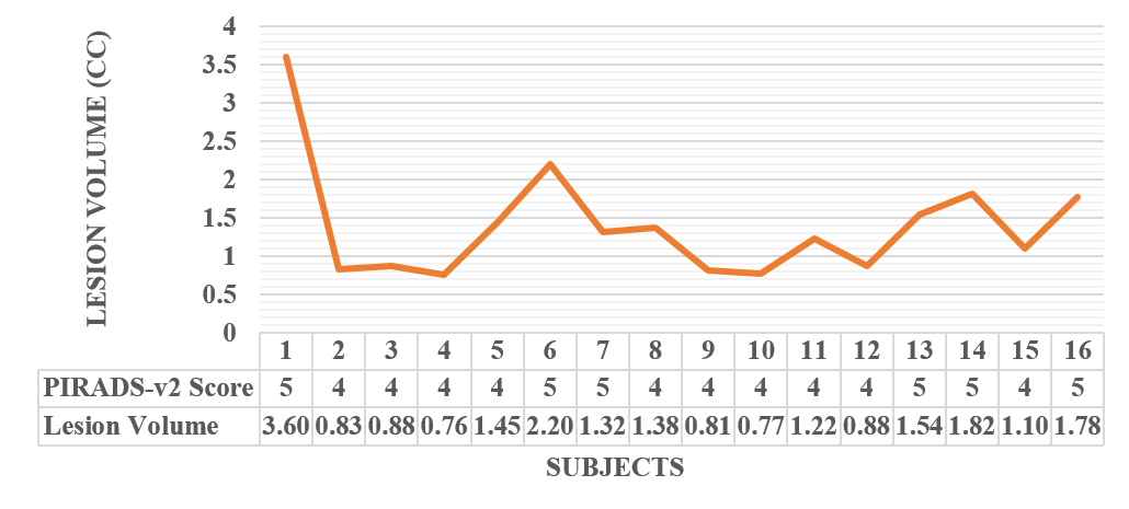
The patient characteristics, radiologist PIRADS-v2 assessment score, and histopathological results are summarized in Table-1. As per radiologist PIRADS-v2 score, of total 16 patients, there were 9 patients with score 4 and 7 patients with score 5. According to Gleason score (GS), 3 patients have highly significant cancer (GS≥7) and 13 patients have intermediate significant cancer (GS<7). Our proposed methodology based PIRADS-v2 assessment shows that out of 16, only one patient is incorrectly classified as PIRADS-v2 score 4, compared to radiologist PIRADS-v2 assessment (sensitiviy,85.71%; specificity, 100%; accuracy,93.75%). When we compared our results to histopathological-score, 3 patients are correctly classified as GS≥7 and PIRADS-v2 score 5; 10 patients are correctly classified as GS<7 and PIRADS-v2 score 4, but 3 patients with GS<7 are incorrectly classified as PIRADS-v2 score 5 (sensitivity,100%; specificity,77%;accuracy,81.25%). The performance of proposed methodology is shown in Table-2.Our study found that the lesion-volume is more than 1.30cc in PIRADS-v2 score 5, which indicates high-grade cancer compare to PIRADS-v2 score 4. The variation of lesion volume with PIRADS-v2 scores is shown in Figure-2. PSA-density is measured by (PSA/Prostate volume)and compared to histopathological PSA-density for verification (Figure-3).
Discussion
In the literature few studies so far evaluate the new guidelines of PIRADS-v2 for diagnosis of PCa; however, they report good diagnostic accuracy, pooled-sensitivity,85%(78-91%), and pooled-specificity,71%(60-80%).7 Our proposed methodology have observed relatively better sensitivity(≥85%),specificity(≥77%),and accuracy(≥81%) compared to radiological PIRADS-v2 assessment and histopathological-score. Lesion-volume is an important finding of this study. This is more than 0.5cc in all patients, which show that all patients have clinically significant cancer and able to easily distinguish between a high and very-high score of cancer. PSA-density has been measured using mpMRI findings which is overestimated as compared to histopathological PSA-density, possibly due to our conservative approach. In future study we will test and validate this approach in a large number of patients towards improving the accuracy of proposed framework.Conclusion
The proposed framework make detection and PIRADS-v2 assessment of PCa using mpMRI more objective and showing good agreement with manual radiologist PIRADS-v2 assessment, which will help radiologists by making the process time-efficient.Acknowledgements
This work is supported by IIT Delhi, India and AIIMS New-Delhi, India. We thanks to Dr Vijay Kubihal for providing clinical inputs. DS was supported with the research fellowship fund from Ministry of Human Resource Development, Government of India.References
1. Ferlay J, Soerjomataram I, Dikshit R, et al. Cancer incidence and mortality worldwide: sources, methods and major patterns in GLOBOCAN 2012. International Journal of Cancer. 2015; 136(5):E359-E386.
2. Boesen L, Multiparametric MRI in detection and staging of prostate cancer. Dan Med J. 2017; 64(2): B5327.
3. Weinreb JC, Barentsz JO, Choyke PL, et al. PI-RADS Prostate Imaging-Reporting and Data System: 2015, Version 2. European Association of Urology. 2016; 69:16-40.
4. Lawrence M, Gallagher FA, Barrett T, et al. Preoperative 3-T diffusion-weighted MRI for the qualitative and quantitative assessment of extracapsular extension in patients with intermediate- or High-Risk Prostate Cancer. American Journal of Roentgenology. 2014; 203(3):280-286.
5. Chan TF, Vese LA, Active contours without edges, IEEE transactions on image processing. 2001; 10(2) :266-277
6. Khalvati F, Salmanpour A, Rahnamayan S, et al. Sequential registration-based segmentation of the prostate gland in MR image volumes. Journal of digital imaging, 2016; 29(2):254–263.
7. Zhang ZX, Yang J, Zhang CZ et al. The value of magnetic resonance imaging in the detection of prostate cancer in patients with previous negative biopsies and elevated prostate-specific antigen levels: A meta-analysis. Acad Radiol. 2014;21:578–89.
Figures