1831
Gland and Zonal Segmentation of Prostate using Diffusion-Weighted MR Imaging1Centre for Biomedical Engineering, Indian Institute of Technology Delhi, New Delhi, India, 2Department of NMR, All India Institute of Medical Sciences Delhi, New Delhi, India, 3Department of Radiology, All India Institute of Medical Sciences Delhi, New Delhi, India, 4Department of Biomedical Engineering, All India Institute of Medical Sciences Delhi, New Delhi, India
Synopsis
Accurate segmentation of the prostate gland and its zones is a challenging task due to the high variability of prostatic anatomic structures. Gland and zonal segmentation of prostate is a useful tool for computer-aided diagnosis of prostate cancer because characteristics of cancer in prostate zones differ significantly. In this study, we used an active contour model for prostate gland segmentation and atlas-based approach for prostate zonal segmentation in diffusion-weighted MR imaging. We have assessed the performance of segmentation methods using different similarity parameters. The proposed methods are highly robust and show relatively good performance compared to previously reported work.
Introduction
Prostate cancer (PCa) is the second most common cancer in men worldwide.1 Multiparametric magnetic resonance imaging (mpMRI) has been developed as an accurate diagnostic tool for the detection of PCa.2 There are distinct glandular regions in prostate glands, consisting of three zones: central zone (CZ),transition zone (TZ) and peripheral zone (PZ). However, CZ and TZ are not distinguishable on MR images and is referred to as TZ. The Prostate Imaging Reporting and Data System version 2 (PI-RADS v2) is a standardized scoring system for reporting of mpMRI of the prostate. The PI-RADS v2 has different algorithms for scoring of PZ and TZ of the prostate as tumors can appear different in these zones.3 Thus, it is important to subsegment the prostate zones in order to use any quantitative analysis methodology. DWI provides superior detection and visualization of PCa.4 Therefore, using DWI for segmentation is a better step, which will be useful in the computer-aided diagnosis (CAD) of PCa. The objective of this study is to optimize the segmentation algorithms for accurate segmentation of prostate gland and its zones.Methods
MRI Data Acquisition: A retrospective dataset of MRI from 18 patients of PCa was used in this study. All prostate mpMRI examinations were performed on a 3T MRI system (Ingenia, Philips, Netherlands) using an external phased array body coil. MRI sequences included axial turbo spin-echo (TSE) T2W (TR/TE=3715/100ms; slice-thickness=3mm; field of view (FOV)=160×160mm2; matrix-size=400×400) and echo-planar DWI (TR/TE=5521/75ms; FOV=177.6×177.6mm2; matrix-size= 176×176; slice-thickness=4mm; with four b-values of 0,500,800 and 1500s/mm2).
Prostate Gland Segmentation: Low b-value (b=0 s/mm2) DWI were used for segmentation of prostate gland as it has better signal to noise ratio and contrast compared to surrounding tissues against higher b-values. The proposed method is based on the active-contour model developed by Chan and Vese.5 The steps of prostate gland segmentation are described in Figure 1.
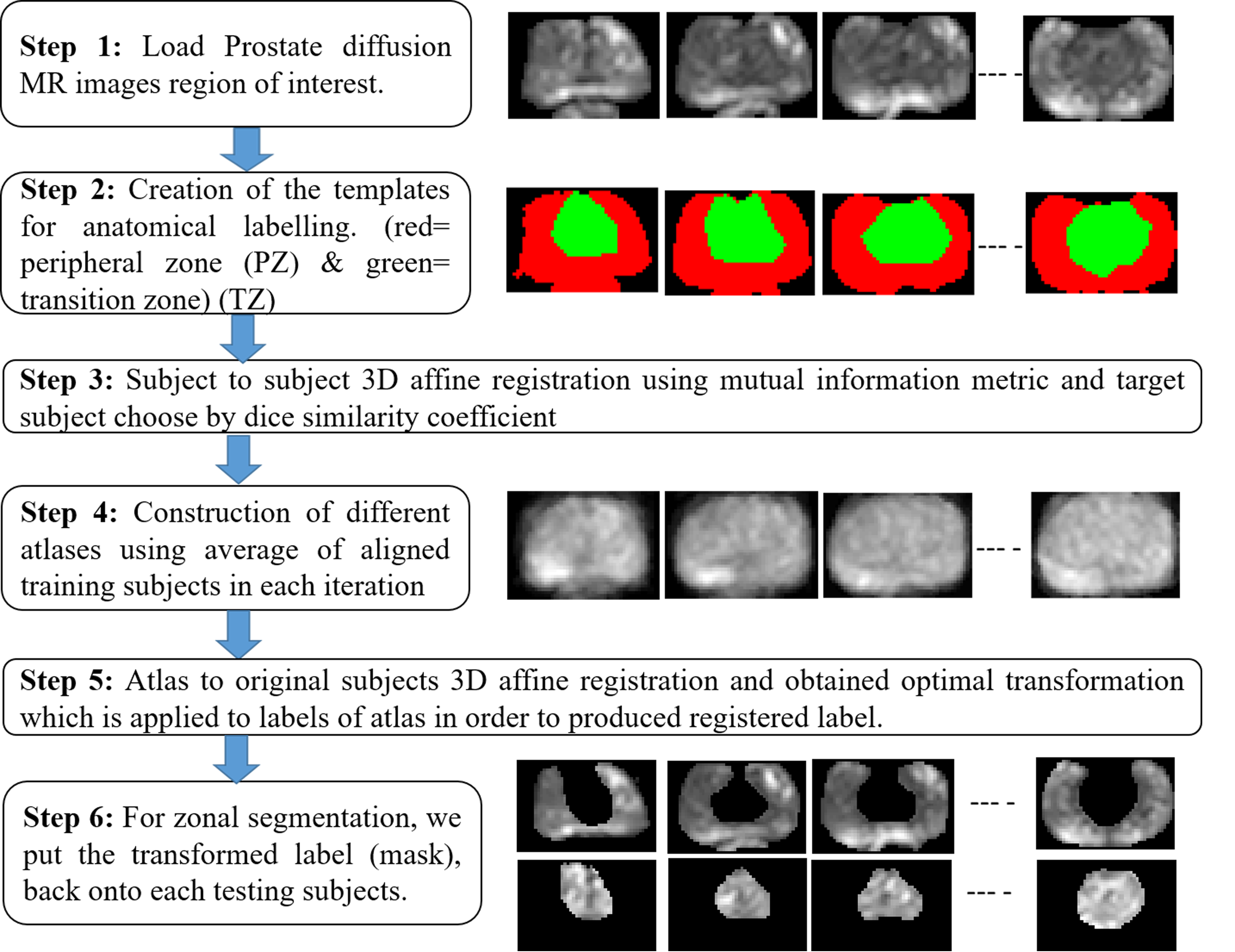
Sub-Segmentation of Prostate to Zonal Segments: The proposed method is based on an atlas-based approach inspired by sequential registration-based segmentation.6 It has two major components; i) registration aiming to align the target image (atlas) and ii) segmentation. The steps of atlas-based zonal segmentation are described in Figure 2.
Data Processing: The atlas-based segmentation method has been assessed with cross-validation. The total dataset was divided into training-set (12 subjects) and testing-set (6 subjects). The training dataset was labeled with manual masking of PZ and TZ. A total of 12 iterations were performed on both training and testing sets. In each iteration, atlases have been constructed by training subjects, and test subjects have been used for validation of zonal segmentation. Data were processed using in-house build algorithms with MATLAB R2017a. Manual segmentation of prostate, PZ, and TZ was performed by an expert radiologist (with more than 10 years of expertise in prostate-imaging) and used as a clinical standard for calculating the performance of segmentation in terms of Dice similarity coefficient (DSC), Jaccard-coefficient, and accuracy.
Results
The average accuracy for the whole prostate gland segmentation of 18 subjects was 99.42± 0.32% with an average DSC and Jaccard-coefficient of 91.06±3.48% and 83.60±5.38% respectively. Using atlas-based technique for segmentation of zones within prostate had an accuracy, DSC and Jaccard-coefficient of 88.60±0.64%, 86.95±1.18%, and 77.41±1.56% respectively for PZ, and for TZ was 93.34±0.70%, 85.10±0.95%, and 74.45±1.36% respectively.Discussion
In this study, we have investigated the abilities of Chen-Vese active contour method for the whole prostate gland segmentation and developed an automated atlas-based prostate zonal segmentation approach for sub-segmentation of the prostate into PZ and TZ using diffusion MR imaging. Compared to the previously published studies, the current one brings three novelties. First, the proposed method operates only on DWI sequence while the most previous methods require multiparametric data 7,8; our results are comparable to the literature. Second, here we have applied morphological processing step to refine the prostate segmentation result; improving the accuracy. The third consists of the development of probabilistic atlas for segmentation of two zones in the prostate. As previously reported, 9-12 mostly the segmentation work has been performed with pooled DSC values of 86% for the prostate gland, 69% for PZ, and 81% for TZ segmentations, respectively. Our methodology achieved relatively better segmentation performance (91% for prostate, 87% for PZ, 85% for TZ) for both whole prostate and its zones using DWI.Conclusion
An active-contour and probabilistic atlas-based methodology for segmentation of prostate and its zones have been developed using DWI. The proposed methods are accurate and easy to implement. This algorithm can be used as a pre-processing step for CAD algorithms or automated PI-RADS classification of PCa in future work.Acknowledgements
This work is supported by IIT Delhi, India and AIIMS New-Delhi, India. We thanks to Dr Vijay Kubihal for providing clinical inputs. DS was supported with the research fellowship fund from Ministry of Human Resource Development, Government of India.References
1. Ferlay J, Soerjomataram I, Dikshit R, et al. Cancer incidence and mortality worldwide: sources, methods and major patterns in GLOBOCAN 2012. International Journal of Cancer. 2015;136(5):E359-E386.
2. Hoeks CM, Barentsz JO, Hambrock T, et al. Prostate cancer: multiparametric MR imaging for detection, localization, and staging. Radiology. 2011;261:46–66.
3. Weinreb JC, Barentsz JO, Choyke PL, et al. PI-RADS Prostate Imaging – Reporting and Data System: 2015, Version 2. European Urology. 2016;69(1):16-40.
4. Kim CK, Park BK, Kim B, et al. Diffusion-weighted MRI at 3 T for the evaluation of prostate cancer. AJR Am J Roentgenol. 2010;194:1461–1469.
5. Chan TF, Vese LA, Active contours without edges, IEEE transactions on image processing. 2001; 10(2):266-277.
6. Khalvati F, Salmanpour A, Rahnamayan S, et al. Sequential registration-based segmentation of the prostate gland in MR image volumes. Journal of digital imaging, 2016; 29(2):254–263.
7. Guo Y, Ruan S, Walker P, et al. Prostate cancer segmentation from multiparametric MRI based on the fuzzy Bayesian model, IEEE 11th International Symposium on Biomedical Imaging (ISBI), Beijing, 2014;866-899.
8. Makni N, Iancu A, Colot O, et al. Zonal segmentation of prostate using multispectral magnetic resonance images. Med Phys. 2011;38(11):6093–6105.
9. Qiu W, Yuan J, Ukwatta E, et al. Dual optimization based prostate zonal segmentation in 3D MR images. Medical Image Analysis. 2014;18(4):660-673.
10. Zhang J, Baig S, Wong A, et al. A local ROI-specific atlas-based segmentation of prostate gland and transitional zone in diffusion MRI, J.Comput.Vision Imaging Syst. 2016;2(1).
11. Clark T, Zhang J, Baig S, et al. Fully automated segmentation of prostate whole gland and transition zone in diffusion-weighted MRI using convolutional neural networks. J Med Imaging. 2017;4(4):041307.
12. Toth R, Ribault J, Gentile J, et al. Simultaneous segmentation of prostatic zones using active appearance models with multiple coupled levelsets. Comput Vis Image Underst. 2013;117(9):1051–1060.
Figures