1823
Diffusion-Weighted Imaging for Predicting Lymph Node Metastasis in Endometrial Cancer: Added Values of Computer-Aided Segmentation and Radiomic Machine Learning1Medical Imaging and Intervention, Chang Gung Memorial Hospital, Taoyuan, Taiwan, 2Clinical Trial Center, Chang Gung Memorial Hospital, Taoyuan, Taiwan
Synopsis
We aim to investigate added values of computer-aided segmentation and radiomic machine learning based on diffusion-weighted magnetic resonance (MR) imaging for predicting nodal metastasis in endometrial cancer. Decision-tree machine learning comprised the apparent diffusion coefficient (ADC), whole tumor volumetric and lymph nodes (LNs) segmentations, MR morphological measurement, and relevant clinical parameters. We concluded that a combination of clinical and MR radiomics generates a prediction model for LNmetastasis in endometrial cancer, with diagnostic performance surpassing the conventional ADC and size criteria.
Purpose
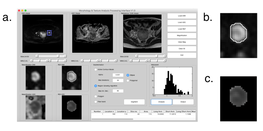
To investigate added values of computer-aided segmentation and radiomic machine learning based on diffusion-weighted magnetic resonance (MR) imaging for predicting nodal metastasis in endometrial cancer.Methods
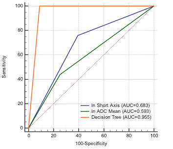
This prospective study included 119 women with endometrial cancer who underwent MRI before surgery during July 2010–June 2015. Decision-tree machine learning comprised the apparent diffusion coefficient (ADC), whole tumor volumetric and lymph nodes (LNs) segmentations, MR morphological measurement, and relevant clinical parameters. Areas under the receiver operating characteristics curve (AUCs) were used to compare diagnostic performances of the decision-tree model, mean ADC criteria, and LNshort-axis diameter.Results
For decision-tree modeling, 106 MRI features and 7 clinical factors were extracted. A decision-tree model was constructed based on grade (1 vs2 and 3), CA125 level (cutoff, 190 U/mL), short-axis diameter of LN(cutoff, 1.8 mm), 75th percentile and minimum ADC of tumor (cutoff, 0.8 × 10⁻³and 0.4 × 10⁻³ mm2/s, respectively), and skewness of relative ADC of LNto tumor (cutoff, 1.3). The sensitivity, specificity, accuracy, positive predictive value, and negative predictive value for the decision-tree model were 100%, 91.1%, 92.0%, 56.8%, and 100%, respectively. The AUC of the decision-tree model was 0.96—significantly higher than the mean ADC (AUC = 0.59, P< 0.0001) and LNshort-axis diameter (AUC = 0.68) criteria (both P< 0.0001).Conclusion
In the present study, we combine all available clinical and MRI parameters to compose an actionable radiomics prediction model using a machine learning method—decision tree analysis. A major advantage of decision tree analysis is the easily interpretable form of the tree using binary splitting rule, which well balances between model accuracy and model simplicity or interpretability, and the familiarity with the modeling techniques of the end user.
We conclude that a combination of clinical and MR radiomics generates a prediction model for LN metastasis in endometrial cancer, with diagnostic performance surpassing the conventional ADC and size criteria.
Acknowledgements
Supported by Chang Gung Medical Foundation grant CIRPG3E0022, CPRPG3G0022, National Science Council (Taiwan) MOST 104-2314-B-182A-095-MY3. Chang Gung IRB 104-8300, 201701215B0.References
Lin G, Ho KC, Wang JJ, et al. Detection of lymph node metastasis in cervical and uterine cancers by diffusion-weighted magnetic resonance imaging at 3T. J Magn Reson Imaging. 2008;28(1):128-35.
Rechichi G, Galimberti S, Oriani M, Perego P, Valsecchi MG, Sironi S. ADC maps in the prediction of pelvic lymph nodal metastatic regions in endometrial cancer. European radiology. 2013;23(1):65-74.
Comparison of retrospective PET and MRI-DWI (PET/MRI-DWI) image fusion with PET/CT and MRI-DWI in detection of cervical and endometrial cancer lymph node metastases. La Radiologia medica. 2016;121(7):537-45.
Figures