1817
Multiregional radiomics features from multiparametric MRI for prediction of lymphovascular invasion in rectal cancer1Department of Radiology, The First Hospital of Jilin University, changchun 130021, China, 2Philips Healthcare, Beijing, China
Synopsis
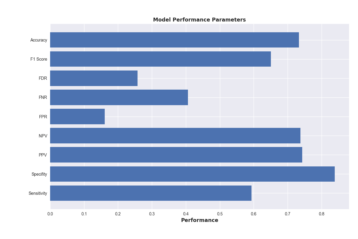
The presence of lymphovascular invasion (LVI) is thought to indicates an increased risk for progressive disease in rectal cancers according to the National Comprehensive Cancer Network (NCCN) Guidelines. Here, we developed and validated a radiomics model for prediction of LVI in rectal cancer based on pre-treatment MRI. The Ridge Classifier have the best prediction accuracy score( 73.3%), its specificity, sensitivity and F1 score are 83.9%, 59.4 % and 65.1 %, respectively. So, the radiomics features from MRI of rectal cancer is a useful tool for predicting LVI preoperatively and has marked discrimination accuracy.
Introduction
One third of Colorectal cancer (CRC) are located in rectum.1 The presence of lymphovascular invasion (LVI) is thought to indicates an increased risk for progressive disease in rectal cancers according to the National Comprehensive Cancer Network (NCCN) Guidelines.2 And the LVI is also considered to be an important biomarker for personalized treatment therapies. 1, 3, 4, 5 The presence of LVI is very important for prognosis and personalized treatment plan in rectal cancer. 1, 3, 4, 5 Rectal magnetic resonance (MR) imaging plays a vital role in the pre-procedure assessment of primary rectal tumor, which is noninvasively. Radiomics, the high-throughput mining of quantitative image features from standard-of-care medical imaging that enables data to be extracted and applied within clinical-decision support systems to improve diagnostic, prognostic, and predictive accuracy, is gaining importance in cancer research.6 We investigated the relationships between the lymphovascular invasion and MR image radiomics features, to evaluate the LVI pre-procedure by MR images.Methods
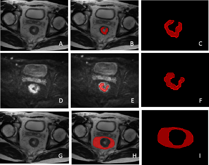
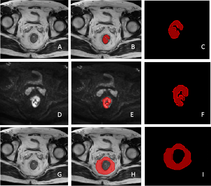
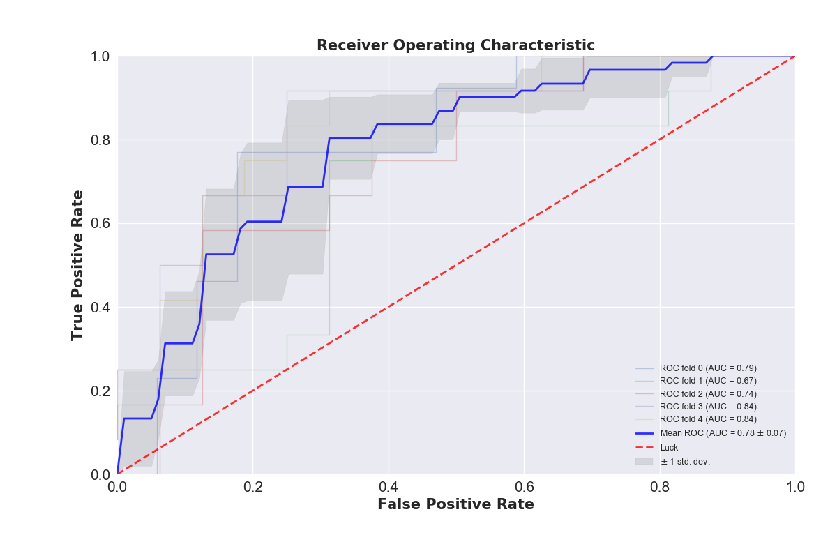
A total of 142 rectal cancer patients who underwent surgical treatment with pathological results between Jan. 2016 and Jan. 2018 were included in this study. All the patients underwent rectal MRI (T2-weighted and diffusion-weighted imaging) scan pre-surgery. 61 patients were diagnosed with LVI. Because the lymphovascular invasion emerges on both primary tumor site and the mesorenctal region, we defined the regions of interest (ROIs) as follows: a. the volume of the whole primary tumor, which were created manually based on T2WI and DWI; b. the volume of mesorectal on T2WI. We use three different series of region of interest (ROI) in Radiomics feature calculation, including lesions of tumor and mesorectum on T2 image and lesions of tumor on DWI image drawn by one radiologist using IntelliSpace Discovery (Philips, Best, the Netherlands) and analyzed by Philips Radiomics Tool (Philips Healthcare, China, the core feature calculation is based on pyRadiomics). For each ROI, a total of 1227 three-dimensional (3D) based radiomic features were extracted. These radiomic features quantified tumor characteristics using tumor size and shape, intensity statistics, and texture. For each patient, we integrated all of the 3681 Radiomics features from three ROIs together. In the following feature dimension reduction analysis, we used Pearson correlation, hierarchical cluster analysis and principal component analysis (PCA) to select the key features. In modeling stage, we investigated 19 classification methods (including Passive Aggressive Classifier, Perceptron, Ridge Classifier, SGD Classifier, Logistic Regression, AdaBoost Classifier, Bagging Classifier, Extra Trees Classifier, Gradient Boosting Classifier, Random Forest Classifier, K Neighbors Classifier, Support Vector Classifier, Decision Tree Classifier, Linear Discriminant Analysis, Quadratic Discriminant Analysis, MLP Classifier, XGB Classifier, Extra Tree Classifier, Gaussian Process Classifier) for training and prediction. These models were trained on the training cohort and their performance was evaluated on the cross-validation cohort using the area under ROC curve (AUC).Result and discussion
Among the 19 machine learning models, the Ridge Classifier is found to give the best prediction accuracy score, the mean value of which is 73.3%. Meanwhile, the mean specificity, sensitivity and F1 score are 83.9%, 59.4 % and 65.1 %, respectively (Figure 3). We developed and validated a radiomics model for the preoperative individualized prediction of LVI in patients with rectal cancer with high accuracy, specificity and sensitivity.Conclusion
Multiparametric radiomics features from multiparametric MRI of rectal cancer help to evaluate the LVI by MR Images.
Acknowledgements
No acknowledgement found.References
1. Glimelius B, Tiret E, Cervantes A, et al. Rectal cancer: ESMO clinical practice guidelines for diagnosis, treatment and follow-up. AnnOncol 2013;24:81–8.
2. NCCN Clinical Practice Guidelines in Oncology (NCCN Guidelines) in Colon Cancer (Version 1.2017). Available at: http://wwwnccnorg/professionals/physician_gls/f_ guidelinesasp. 2017.
3. Benson AB, Schrag D, Somerfield MR, et al. American society of clinical oncology recommendations on adjuvant chemotherapy for stage II colon cancer. J Clin Oncol 2004;22:3408–19.
4. Petersen VC, Baxter KJ, Love SB, et al. Identification of objective pathological prognostic determinants and models of prognosis in Dukes' B colon cancer. Gut 2002;51:65–9.
5. Quah HM, Chou JF, Gonen M, et al. Identification of patients with high-risk stage II colon cancer for adjuvant therapy. Dis Colon Rectum 2008;51:503–7.
6.Lambin P, Leijenaar RTH, Deist TM, et al. Radiomics: the bridge between medical imaging and personalized medicine. Nature reviews Clinical oncology. 2017;14(12):749-62.
Figures