1815
Rectal cancer: preoperative prediction of perineural invasion by machine learning modeling of multiregional radiomics features from multiparametric MRI1Department of Radiology, The First Hospital of Jilin University, changchun 130021, China, 2Philips Healthcare, Beijing, China
Synopsis
Defined by tumor invasion of nervous structures and nerve sheaths, the presence of perineural invasion (PNI) is thought to indicate an increased risk for progressive disease in rectal cancers. Here, we developed and validated a radiomics model for individualized prediction of PNI in rectal cancer based on pre-procedure MRI. The Ridge Classifier is found to have the best prediction accuracy score (80.8%), its specificity, sensitivity and F1 score are 90.5%, 60.4%, and 67.0%, respectively. So, the radiomics features from MRI of rectal cancer is a useful tool for predicting PNI preoperatively and has marked discrimination accuracy.
Introduction
There are more than 100,000 people diagnosed with rectal cancer worldwide every year1. Defined by tumor invasion of nervous structures and nerve sheaths, the presence of perineural invasion (PNI) is thought to indicate an increased risk for progressive disease in rectal cancers according to the National Comprehensive Cancer Network (NCCN) Guidelines 1,2 And the PNI is also considered to be an important biomarker for personalized treatment therapies .3-6The presence of perineural invasion (PNI) is very important for prognosis and personalized treatment plan in rectal cancer.3-6 Rectal magnetic resonance (MR) imaging plays a vital role in the pre-procedure assessment of primary rectal tumor, which is noninvasively compared with other modalities such as preoperative biopsy and serum test. 7, 8 Currently,“radiomics”has enabled the extraction of more detail quantitative features from conventional MRI. 7-10 Whether there exist an association between the pathologic feature of PNI and radiomics features of MR images in rectal cancer, has not been studied, to the best of our knowledge. Therefore, the aim of this study was to develop and validate a radiomics prediction model based on pre-procedure MRI (T2-weighted and diffusion-weighted imaging), to predict of perineural invasion (PNI) in rectal cancer.Methods
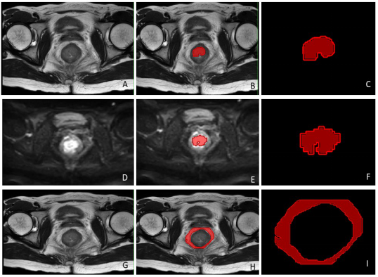
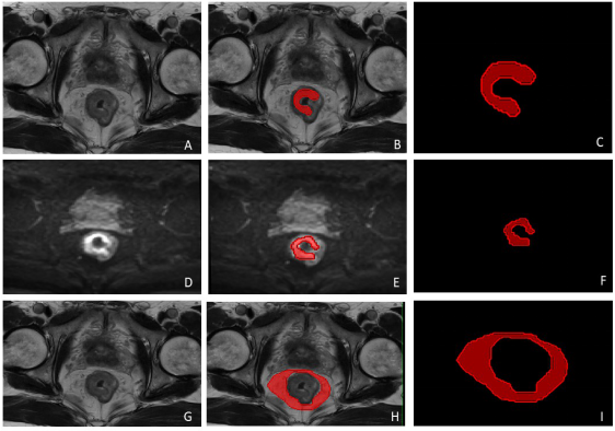
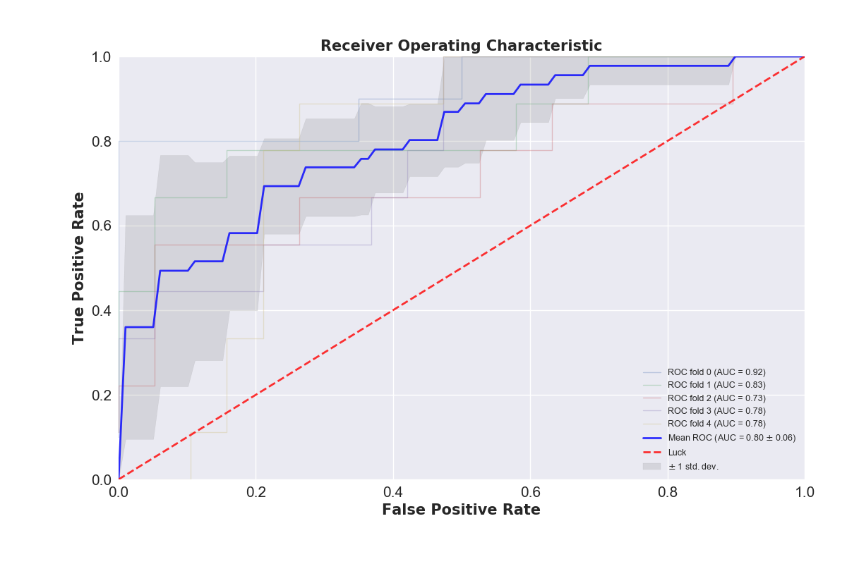
A total of 142 rectal cancer patients who underwent radical surgical resection with pathological results between Jan. 2016 and Jan. 2018 were included in this study. All the patients underwent rectal MRI (T2-weighted and diffusion-weighted imaging) scan pre-surgery. Multivariate models were trained on the training cohort and their performance was evaluated on the 5-fold cross-validation cohort using the area under ROC curve (AUC), accuracy, specifity and sensitivity. The regions of interest (ROIs) were the volume of the whole primary tumor, which were created manually based on T2WI and DWI data slice by slice. We use three different series of region of interest (ROI) in Radiomics feature calculation, including lesions of tumor and mesorectum on T2 image and lesions of tumor on DWI image drawn by one radiologist using IntelliSpace Discovery (Philips, Best, the Netherlands) and analyzed by Philips Radiomics Tool (Philips Healthcare, China, the core feature calculation is based on pyRadiomics). For each ROI, a total of 1227 three-dimensional (3D) based radiomic features were extracted. These radiomic features quantified tumor characteristics using tumor size and shape, intensity statistics, and texture. For each patient, we integrated all of the 3681 Radiomics features from three ROIs together. In the following feature dimension reduction analysis, we used Pearson correlation, hierarchical cluster analysis and principal component analysis (PCA) to select the key features. In modeling stage, we investigated 19 classification methods (including Passive Aggressive Classifier, Perceptron, Ridge Classifier, SGD Classifier, Logistic Regression, AdaBoost Classifier, Bagging Classifier, Extra Trees Classifier, Gradient Boosting Classifier, Random Forest Classifier, K Neighbors Classifier, Support Vector Classifier, Decision Tree Classifier, Linear Discriminant Analysis, Quadratic Discriminant Analysis, MLP Classifier, XGB Classifier, Extra Tree Classifier, Gaussian Process Classifier) for training and prediction. These models were trained on the training cohort and their performance was evaluated on the cross-validation cohort using the area under ROC curve (AUC).Result and discussion
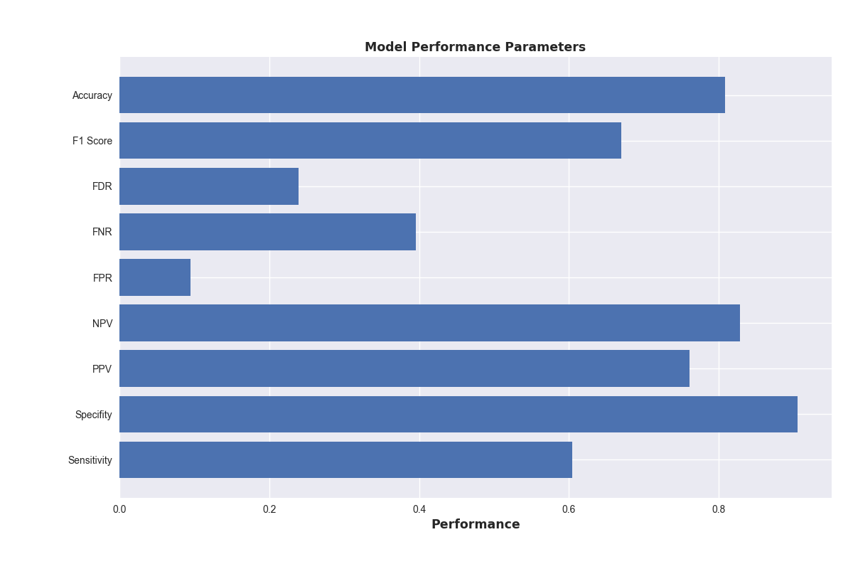
Among the 19 machine learning models, the Ridge Classifier is found to have the best prediction accuracy score(80.8%), its specificity, sensitivity and F1 score are 90.5%, 60.4%, and 67.0%, respectively (Figure 4). We developed and validated a diagnostic, radiomics method for the preoperative individualized predictionof PNI in patients with rectal cancer with high accuracy, specificity and sensitivity.Conclusion
Multiparametric radiomics features from multiparametric MRI of rectal cancer is a useful tool for predicting Perineural Invasion (PNI) preoperatively and has marked discrimination accuracy.Acknowledgements
No acknowledgement found.References
1. Glimelius B, Tiret E, Cervantes A, et al. Rectal cancer: ESMO clinical practice guidelines for diagnosis, treatment and follow-up. AnnOncol 2013;24:81–8.
2. Liebig C, Ayala G, Wilks JA, et al. Perineural invasion in cancer: a review of the literature. Cancer 2009;115:3379-91.
3. Liebig C, Ayala G, Wilks J, et al. Perineural invasion is an independent predictor of outcome in colorectal cancer. J Clin Oncol 2009;27:5131-7.
4. Huh JW, Kim HR, Kim YJ. Prognostic value of perineural invasion in patients with stage II colorectal cancer. Ann Surg Oncol 2010;17:2066-72.
5. Suzuki T, Suwa K, Ogawa M, et al. Adjuvant chemotherapy for the perineural invasion of colorectal cancer. J Surg Res 2015;199:84-9.
6. Yang Y, Huang X, Sun J, et al. Prognostic value of perineural invasion in colorectal cancer: a metaanalysis.J Gastrointest Surg 2015;19:1113-22.
7. Garcia-Figueiras R, Baleato-Gonzalez S, Padhani AR, et al. Advanced Imaging Techniques in Evaluation of Colorectal Cancer. Radiographics : a review publication of the Radiological Society of North America, Inc. 2018;38(3):740-65.
8. Horvat N, Veeraraghavan H, Khan M, et al. MR Imaging of Rectal Cancer: Radiomics Analysis to Assess Treatment Response after Neoadjuvant Therapy. Radiology. 2018;287(3):833-43.
9. Lambin P, Leijenaar RTH, Deist TM, et al. Radiomics: the bridge between medical imaging and personalized medicine. Nature reviews Clinical oncology. 2017;14(12):749-62.
10. Limkin EJ, Sun R, Dercle L, et al. Promises and challenges for the implementation of computational medical imaging (radiomics) in oncology. Annals of oncology : official journal of the European Society for Medical Oncology. 2017;28(6):1191-206.
Figures