1812
Locally Advanced Rectal Cancer: The Value of Intravoxel Incoherent Motion Imaging and Diffusion Kurtosis Imaging in Assessing Tumor Response to Neoadjuvant Chemoradiotherapy1West China Hospital, Chengdu, China
Synopsis
This study combined IVIM and DKI sequences in assessing tumor response to neoadjuvant chemoradiotherapy in locally advanced rectal cancer. Although both IVIM and DKI model derived parameters in our study could help to identify complete responders, ADC value was found to outperform both IVIM and DKI paramters in selecting complete responders.
Purpose
To investigate the role of intravoxel incoherent motion imaging (IVIM) and diffusion kurtosis imaging (DKI) in assessing tumor response to neoadjuvant chemoradiotherapy (CRT) in locally advanced rectal cancer (LARC).Methods and Materials
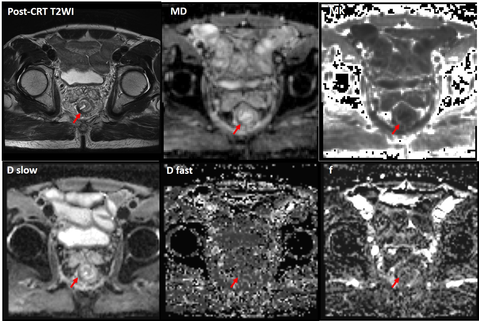
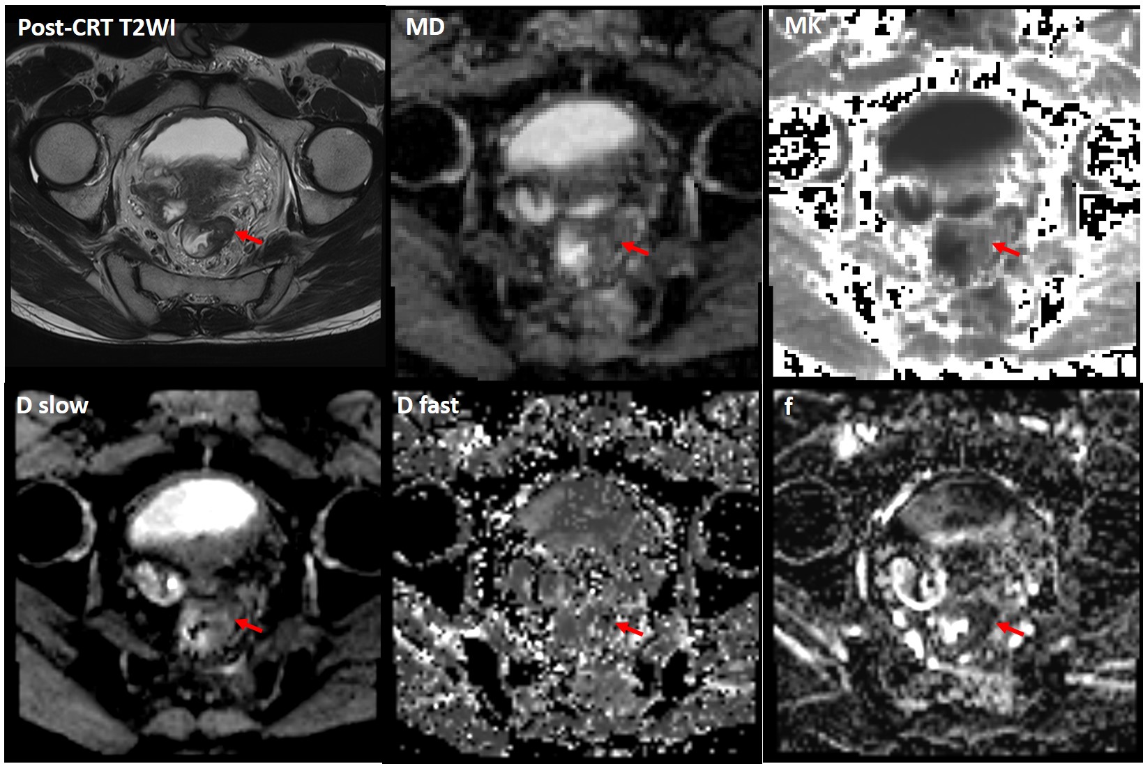
Between July 2017 to Oct 2018, 32 LARC patients (cT3/4 or N+) were enrolled in this prospective study, and underwent pre- and post-CRT rectal MRI on a 3.0 T MRI scanner, including IVIM and DKI sequences with 12 b values. They all received neoadjuvant CRT and subsequent surgery. Histopathological tumor regression grade (TRG) of the surgical specimen served as the reference standard. Patients were divided into pCR (TRG0) and non-pCR group (TRG1-3). Mean slow diffusion coefficient (Dslow) (*10-3 mm2/s), fast diffusion coefficient (Dfast) (*10-3 mm2/s), perfusion-related diffusion fraction (f), mean kurtosis (MK), mean diffusion (MD) (*10-3 mm2/s) and monoexponential ADC value (*10-3 mm2/s) were calculated by manually drawing ROIs on three representative slices of primary and residual tumor on pre- and post-CRT b=800 s/mm2 images. ROIs were then copied to images of IVIM and DKI parameters. Independent t test, Mann-Whitney U test, and ROC curves were used for statistical analyses.Results
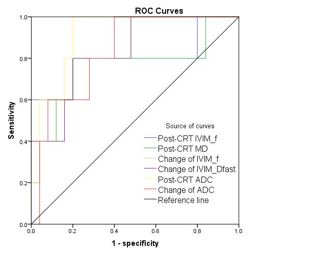
The pCR group (n=6) had a significant higher post-CRT f (0.138±0.036 vs. 0.1±0.023, P=0.003), MD (1.78±0.48 vs. 1.406±0.264, P=0.013) and ADC value (1.495±0.16 vs. 1.234±0.169, P=0.004) than non-pCR group (n=26). Also the change of f (0.038±0.036 vs. -0.005±0.028, P=0.003), Dfast (2.211 vs. -6.2, P=0.007) and ADC value (0.369±0.119 vs. 0.177±0.201, P=0.05) were significant higher in the pCR group after treatment. ROC curves showed that Post-CRT f, MD and ADC value presented AUCs of 0.801, 0.769, 0.929 in selecting pCR. And Changes of f, Dfast and ADC value after treatment all presented AUCs of 0.832 in differentiating complete response. Post-CRT MD and ADC value was negatively correlated with TRG (rs=-0.399 and -0.641, P=0.024 and <0.001).Conclusions
IVIM model derived f, Dfast, and DKI model derived MD value could help to identify pCR after neoadjuvant CRT in LARC. But post-CRT monoexponential ADC value showed the best diagnostic performance in selecting pCR.Acknowledgements
No acknowledgement found.References
1. Smith J J and Garcia-Aguilar J Advances and challenges in treatment of locally advanced rectal cancer. J Clin Oncol, 2015, 33(16):1797-1808.
2. Iima M and Le Bihan D Clinical Intravoxel Incoherent Motion and Diffusion MR Imaging: Past, Present, and Future. Radiology, 2016, 278(1):13-32.
3. Rosenkrantz, Andrew B.; Padhani, Anwar R.; Chenevert, Thomas L.; Koh, Dow‐mu ; De Keyzer, Frederik; Taouli, Bachir; Le Bihan, Denis (2015). "Body diffusion kurtosis imaging: Basic principles, applications, and considerations for clinical practice." Journal of Magnetic Resonance Imaging 42(5): 1190-1202.
Figures