1805
Predictive value of intravoxel incoherent motion imaging (IVIM) and diffusion kurtosis imaging (DKI) in patients with locally advanced cervical cancers treated with neoadjuvant chemoradiotherapy1The Second Hospital of Dalian Medical University, Dalian, China, 2GE Healthcare, Beijing, China
Synopsis
The purpose of this study was to investigate the use of IVIM and DKI to predict the efficacy of neoadjuvant chemoradiotherapy (NACT) in locally advanced cervical cancer. We found that the ADC, D and f values of IVIM and the MK value of DKI were significantly changed before and after NACT. Therefore, these parameters are predictive in cervical cancer treated with NACT.
Purpose
The purpose of this study is to explore the value of incoherent motion diffusion weighted imaging (IVIM) and diffusion kurtosis imaging (DKI) in the evaluation of the efficacy of neoadjuvant chemoradiotherapy (NACT) for cervical cancer.Introduction
Cervical cancer is the second most common cancer among women with the highest mortality rate in female genital malignancies. The routine treatment for advanced cervical cancer is radiotherapy. However, radiotherapy damages the ovarian function and affects the quality of life. For young and middle-aged patients, NACT becomes the trend of treatment for locally advanced cervical cancer (LACC) (FIGO IB2∼ IIB) [1], while the drawback of NACT is that failure of chemotherapy may lead to the delay of cancer treatment. Therefore, it is essential to evaluate the efficacy of NACT in order to avoid delays in exact surgery or radiation treatment [2]. As the cutting-edge functional imaging technologies, IVIM and DKI could provide biological information beyond morphology in terms of molecules and metabolism. Furthermore, both technologies have been applied in the evaluation of therapeutic effect and prognosis of various malignant tumors. To our knowledge, the role of IVIM and DKI in the assessment of the prognosis and efficacy of cervical cancer treated with NACT has not been reported.Materials and Methods
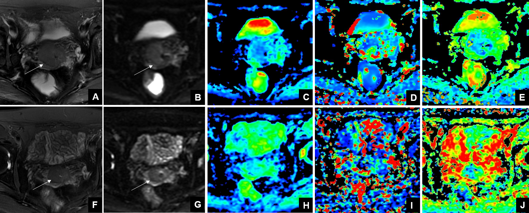
47 females (age 43.1±12.0 years, age range 32∼ 68 years) with histologically proven untreated cervical cancer (FIGO IB2 ∼ IIB) were scheduled to undergo NACT. All the patients underwent routine MRI examination, IVIM and DKI examination before and after NACT on a 3.0T MR station (Discovery MR750W, GE Healthcare, USA). The IVIM examination with 11 b values (0, 20, 50, 100, 150, 200, 400, 800, 1000, 1200 and 1500 s/mm2) and the DKI examination with 3 b values (0, 1000 and 2000 s/mm2 ) in 15 directions. Images analyses were performed on GE AW4.6 workstation by two radiologists. The efficacy of NACT depends on the changes of the tumor size after two weeks of treatment. According to the Response Evaluation Criteria in Solid Tumors (RECIST) [3], we divided the patients into effective group (including complete response and partial response) and ineffective group (including stable disease and progressive disease).Results
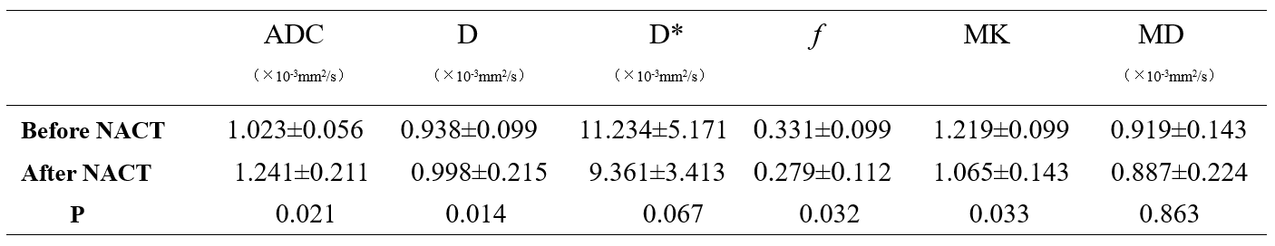
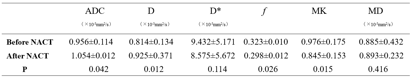
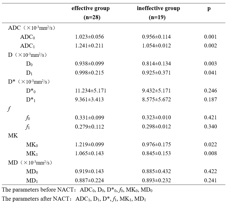
After NACT treatment, the ineffective group consisted of 28 patients and the ineffective group contained of 19 patients. The ADC and D values were significantly higher and the f and MK values were significantly lower after NACT treatment (both in the effective group and ineffective group). No significant difference in the D* and MD values were observed before and after NACT (both in the effective group and ineffective group) as shown in Table 1 and 2. The ADC, D and MK values were significantly higher both before and after NACT in the effective group compared with that of the ineffective group. No significant differences in the D*, f and MD values were observed before and after NACT in the effective group compared with that of the ineffective group (the P value is 0.001 and 0.002 for ADC; 0.003 and 0.041 for D; 0.246 and 0.187 for D*; 0.421 and 0.340 for f; 0.022 and 0.008 for MK; 0.422 and 0.241 for MD) as shown in Table 3.Discussion and Conclusion
RECIST plays an important role in evaluating the short-term response of cervical cancer patients when receiving NACT. Previous reports have shown that the IVIM and DKI parameters have been successfully used to distinguish malignant cervical lesions from benign ones, and the pathological grades of cervical cancers. The results of this study indicate that the parameters of IVIM and DKI are full of potential to predict the efficacy of NACT in cervical cancers. The ADC and D values decrease and the f and MK values increase after NACT, and the ADC, D and MK values are significantly higher in the effective group when compared to that of the ineffective group, either before or after NACT treatment. Since the increased cellular density attributes to the low diffusion values of tumors [4], we conclude that the cervical cancer of the effective group may have lower cellular density compared with that of the ineffective group. This study indicates these diffusion and kurtosis indices from IVIM and DKI, the ADC and D values of IVIM and the MK values of DKI may perform as a predictor of the efficacy of NACT for cervical cancer treatment.Acknowledgements
No acknowledgement found.References
[1] Fu C, Bian D, Liu F, et al. The value of diffusion-weighted magnetic resonance imaging in assessing the response of locally advanced cervical cancer to neoadjuvant chemotherapy[J]. International Journal of Gynecological Cancer, 2012, 22(6): 1037-1043.
[2] Ren Y, Li Y, Liu J. A modified shortened administration schedule for neoadjuvant chemotherapy with irinotecan and cisplatin in locally advanced cervical cancer[J]. International Journal of Gynecological Cancer, 2011, 21(4): 685-689.
[3] Chalian H, Töre H G, Horowitz J M, et al. Radiologic assessment of response to therapy: comparison of RECIST versions 1.1 and 1.0[J]. Radiographics, 2011, 31(7): 2093-2105.
[4] Wang Y C, Hu D Y, Hu X M, et al. Assessing the early response of advanced cervical cancer to neoadjuvant chemotherapy using intravoxel incoherent motion diffusion-weighted magnetic resonance imaging: a pilot study[J]. Chinese medical journal, 2016, 129(6): 665.
Figures