1804
Correlation study between parameters of intravoxel incoherent motion diffusion-weighted imaging and different pathological differentiation of cervical squamous cell carcinoma1anhui provincal hospital, hefei, China
Synopsis
Correlation study between parameters of intravoxel incoherent motion diffusion-weighted imaging and different pathological differentiation of cervical squamous cell carcinoma
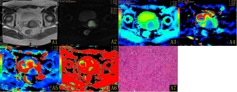
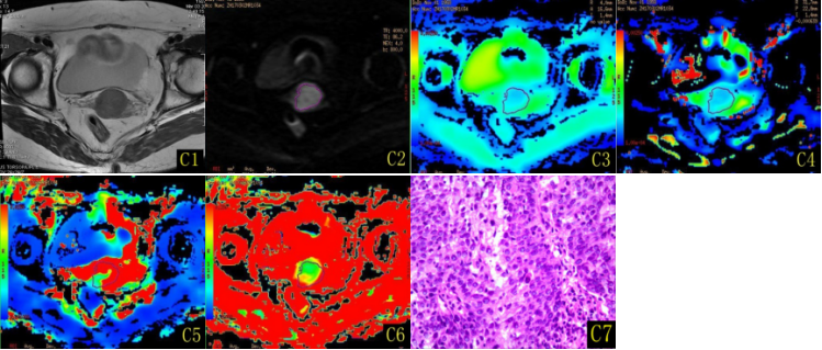
Objective To evaluate the correlation between parameters of intravoxel incoherent motion diffusion-weighted imaging and different pathological differentiation degree of cervical squamous cancer. Methods Retrospective analysis of 87 cases patients of cervical squamous cell cancer confirmed by surgery and pathology in our hospital were included in this study, which fifty cases were in the low differentiation group(Fig A1-A7), twenty six cases were in the middle differentiation group(Fig B1-B7) and eleven cases were highly differentiation(Fig C1-C7),the conventional magnatic resonance imaging and intravoxel incoherent motion diffusion-weighted imaging examinations were performed in all patients, intravoxel incoherent motion diffusion-weighted imaging was performed by using 10 b values(b=0,10,20,50,100,200,400,800,1200,2000s/mm2). ADCstand, pure water molecular diffusion coefficient (D), perfusion diffusion coefficient (D*) and perfution fraction (f) of IVIM - DWI related parameter values were measured with functool software of GE ADW4.5 workstation respectively.Statistical analysis software SPSS 22.0 was used in differential cervical squamous cell carcinoma diffentiation and IVIM - DWI related parameters.The receiver operating characteristic curve(ROC) was used to distinguish the efficiency of differentiation degree of cervical squamous cell carcinoma. Results The ADCstand, D, D*and f values of low differentiation group were (0.72±0.11) ×10-3mm2/s, (0.45±0.067) ×10-3mm2/s, (7.56±3.09) ×10-3mm2/s and (39.55,14.25)%; The ADCstand, D, D*and f values of middle differentiation group were (0.74±0.11)×10-3mm2/s, (0.58±0.036)×10-3mm2/s, (17.19±4.37) ×10-3mm2/s and (24.05,3.73)%; The ADCstand, D, D*and f values of highly differentiation were (0.78±0.073) ×10-3mm2/s, (0.74±0.024) ×10-3mm2/s, (39.18±10.09) ×10-3mm2/s and (18.70,1.60)%; There are statistical significance in the D, D*and f values of inter-groups(P<0.05),but there is no statistical significance in the ADCstand of inter-groups(P=0.19). The D and D* values were positively correlated with the pathological differentiation of squamous cell carcinoma (r was 0.853,0.880), and the f value was moderately negative correlation (r = -0.730). In the diagnosis of low differentiation and high differentiation of cervical squamous cell carcinoma, the D value had the best diagnostic efficiency; In the diagnosis of cervical squamous cell carcinoma, f value has the best diagnostic efficiency. Conclusion The D, D * and f values of IVIM-DWI can non-invasively assess the pathological differentiation degree of cervical squamous cell cancers,It is helpful for the clinican to develop a scientific and individualized treatment plan of cervical cancer, so as to improve the efficacy of it.
Key words Intravoxel incoherent motion; Diffusion weighed imaging; cervical squamous cell carcinoma; pathological histological differentiation
Acknowledgements
No acknowledgement found.References
[1]张庆, 徐香玖, 周星,等. 3.0TMR动态对比增强对宫颈癌组织学特性和临床分期的分析[J]. 临床放射学杂志, 2015, 34(10):1607-1610.
[2]Liu Y, Bai R, Sun H, et al. Diffusion-weighted magnetic resonance imaging of uterine cervical cancer.[J]. Journal of Computer Assisted Tomography, 2009, 33(6):858-862.
[3]Chen J, Zhang Y, Liang B, et al. The utility of diffusion-weighted MR imaging in cervical cancer[J]. European Journal of Radiology, 2010, 74(3):101-6.
[4] Mcveigh P Z, Syed A M, Milosevic M, et al. Diffusion-weighted MRI in cervical cancer[J]. European Radiology, 2008, 18(5):1058-64.
[5] 刘颖, 白人驹. DWI在宫颈癌诊断中的应用价值及其与病理相关性[J]. 临床放射学杂志, 2009, 28(2):225-229.
[6] 何长久, 阳宁静, 董晓蕾,等. 扩散加权成像在宫颈癌诊断中的价值[J]. 实用放射学杂志, 2015(8):1316-1318.[7] Naganawa S, Sato C, Kumada H, et al. Apparent diffusion coefficient in cervical cancer of the uterus: comparison with the normal uterine cervix.[J]. European Radiology, 2005, 15(1):71-78.
[8] 王越. 宫颈癌与正常宫颈的PWI、MRS影像学表现及其临床价值的初步探讨[D]. 大连医科大学, 2014.
[9] Mahon M M, Cox I R, Soutter W P, et al. 1H magnetic resonance spectroscopy of preinvasive and invasive cervical cancer: in vivo-ex vivo profiles and effect of tumor load[J]. Journal of Magnetic Resonance Imaging, 2004, 19(3):356-364.
[10] 邝菲, 颜志平, 冯浩. 3.0T DCE-MRI对宫颈癌的评估价值[J]. 实用放射学杂志, 2016, 32(3)
.[11] Becker A S, Perucho J A, Wurnig M C, et al. Assessment of Cervical Cancer with a Parameter-Free Intravoxel Incoherent Motion Imaging Algorithm [J]. Korean Journal of Radiology, 2017, 18(3):510-518.
[12] Yan Z, Liu J, Liu C, et al. Intravoxel incoherent motion diffusion weighted MRI of cervical cancer — Correlated with tumor differentiation and perfusion[J]. Magnetic Resonance Imaging, 2016, 34(8):1050-1056.
[13] 王欢欢, 周正扬, 朱丽晶,等. IVIM-MRI评估宫颈癌同步放化疗疗效探讨[J]. 中华放射肿瘤学杂志, 2016, 25(10):1100-1105.
[14] Park J J, Kim C K, Park S Y, et al. Assessment of early response to concurrent chemoradiotherapy in cervical cancer: value of diffusion-weighted and dynamic contrast-enhanced MR imaging.[J]. Magnetic Resonance Imaging, 2014, 32(8):993-1000.
[15] Akita A, Shinmoto H, Hayashi S, et al.Comparison of T2 –weighted and contrast-enhanced T1 -weighted MR imaging at 1.5T for assessing the local extend of cervical carcinom[J]. European Radiology,2011,21: 1850-1857.
[16] 刘凤海, 李国策, 刘世凯,等. 3.0 T高分辨力MRI对Ⅱ期宫颈癌精确分期的价值[J]. 临床放射学杂志, 2017, 36(1):85-89
.[17] Fu ZZ,Peng Y,Cao LY,et al. Value of apparent diffusion coefficient( ADC) in assessing radiotherapy and chemotherapy success in cervical cancer[J]. Magnetic Resonance Imaging, 2015,33: 516-524.
[18] Park JJ, Kim CK, Park SY, et al.Assessment of early response to concurrent chemoradiotherapy in cervical cancer: value of diffusionweighted and dynamic contrast-enhanced MR imaging [J]. Magnetic Resonance Imaging,2014,32: 993-1000.
[19] 吴斌, 黄啸, 彭卫军,等. 磁共振扩散加权成像在宫颈癌诊断和疗效预测中的价值[J]. 中华肿瘤杂志, 2014, 36(2):115-119.
[20] Schreuder SM, Lensing R, Stoker J, et al. Monitoring treatment response in patients undergoing chemoradiotherapy for locally advanced uterine cervical cancer by additional diffusion-weighted imaging: A systematic review[J] . J Magn Reson Imaging,2015,42: 572-594.
[21] Loncaster JA, Carrington BM, Sykes JR, Jones AP, Todd SM, Cooper R, et al.Prediction of radiotherapy outcome using dynamic contrast enhanced MRI of carcinoma of the cervix. Int J Radiat Oncol Biol Phys 2002;54:759–67.
[22] Zahra MA, Tan LT, Priest AN, et al.Semiquantitative and quantitative dynamic contrast-enhanced magnetic resonance imaging measurements predict radiation response in cervical cancer [J]. Int J Radiat Oncol Biol Phys, 2009, 74: 766-773.
[23] Jalaguier-coudray A, Villard-mahjoub R, Delouche A, et al. Value of Dynamic Contrast-enhanced and Diffusion-weighted MR Imaging in the Detection of Pathologic Complete Response in Cervical Cancer after Neoadjuvant Therapy: A Retrospective Observational Study.[J]. Radiology, 2017:161299.
[24] Ovreb KM, Ellingsen C, Hompland T, et al. Dynamic contrast-enhanced magnetic resonance imaging of the metastatic potential of tumors: a preclinical study of cervical carcinoma and melanoma xenografts[J].Acta Oncol, 2012,52: 604-611.
[25] Zhu L, Zhu L, Shi H, et al. Evaluating early response of cervical cancer under concurrent chemo-radiotherapy by intravoxel incoherent motion MR imaging.[J]. Bmc Cancer, 2016, 16(1):1-8.
[26] 朱志军.MRI动态增强定量分析宫颈鳞癌微血管通透性价值的研究[J].中国 CT 和MRI杂志,2015,13: 71-73.
Figures