1799
Quantitative 31P magnetic resonance spectroscopy detects insulin-mediated reduction of hepatic ATP content in type 2 diabetic patients1Institute for Clinical Diabetology, German Diabetes Center, Leibniz Institute for Diabetes Research at Heinrich Heine University, Düsseldorf, Germany, 2German Center for Diabetes Research (DZD e.V.), München-Neuherberg, Germany, 3Division of Endocrinology and Diabetology, Medical Faculty, Heinrich Heine University Düsseldorf, Düsseldorf, Germany
Synopsis
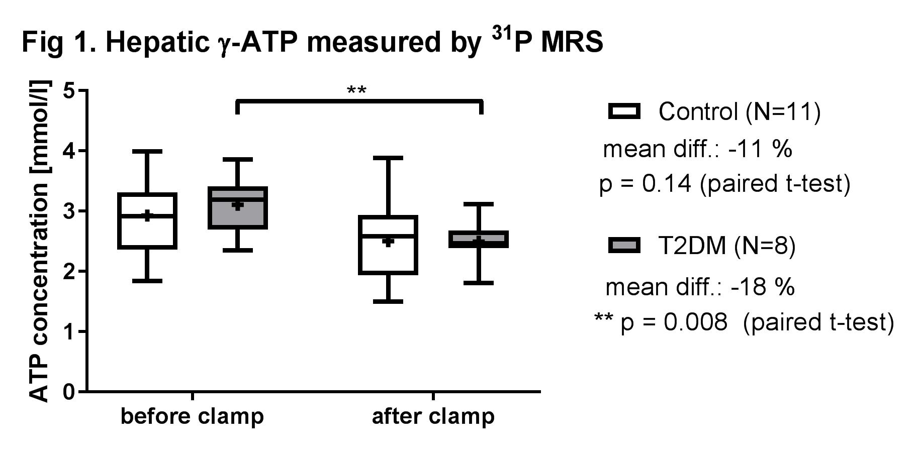
Decreased insulin sensitivity precedes the onset of type 2 diabetes mellitus (T2DM). Hepatic energy metabolism is assumed to be impaired in T2DM with reduced insulin sensitivity. However, little is known about changes of ATP content in response to acutely elevated insulin. Therefore utilizing 31P MRS, we quantified ATP content before and after acute hyperinsulinemia. After a hyperinsulinemic clamp test, hepatic γ-ATP concentrations (ATP) were significantly reduced by 18% (p=0.008) in T2DM but not in controls. This study demonstrated that quantitative 31P MRS allows to monitor acute changes in hepatic ATP concentrations in vivo in various metabolic conditions.
INTRODUCTION
Decreased insulin sensitivity is known to precede the clinical onset of type 2 diabetes mellitus (T2DM). T2DM patients feature lower insulin-stimulated flux through ATP synthesis in skeletal muscle1. Hepatic energy metabolism may be also impaired in T2DM2. However, little is known about changes of hepatic ATP content in response to acutely elevated insulin levels. Thus, the aim of this study was to quantify changes of ATP content employing 31P magnetic resonance spectroscopy (MRS) in T2DM and glucose tolerant humans before and after standardized hyperinsulinemia.METHODS
After consenting to the study approved by the local IRB, 8 T2DM (age: 56.4 ± 7.8 yrs; body mass index, BMI: 29.3 ± 2.3 kg/m²) and 11 glucose tolerant controls with comparable age and BMI (CON, 56.9 ± 8.5 yrs; 29.0 ± 2.1 kg/m²) underwent stepped low and high dose insulin-glucose clamps combined with isotopic dilution of [6,6-2H2] glucose to assess hepatic and peripheral insulin sensitivity at constant blood glucose concentrations at 5 mmol/l as previously published3.
MR Study All MR measurements were performed on a 3-Tesla MR scanner (Achieva X-series, Philips Healthcare, Best, the Netherlands). A 14-cm circular 31P receiver/transmitter RF coil was used for 31P MRS. The built-in body coil was utilized for MR imaging, proton decoupling and NOE as reported4. Transverse and coronal images were acquired to properly localize a 6×6×6 cm3 voxel within the liver for 31P MRS. The acquisition sequence parameters of 31P MRS were TR/NSA/pulse sequence = 6 s/128/3D ISIS with an adiabatic excitation pulse (spectral width = 3000 Hz, data points = 2048). 1H MRS for the liver fat was acquired using a SENSE receive coil. Liver fat content was quantified from non-water-suppressed 1H MR spectra using STEAM and expressed as percent fat relative to water content4,5. All spectra were processed with jMRUI. Final ATP content was quantified in mmol/l after correcting for fat content of each individual4.
RESULTS
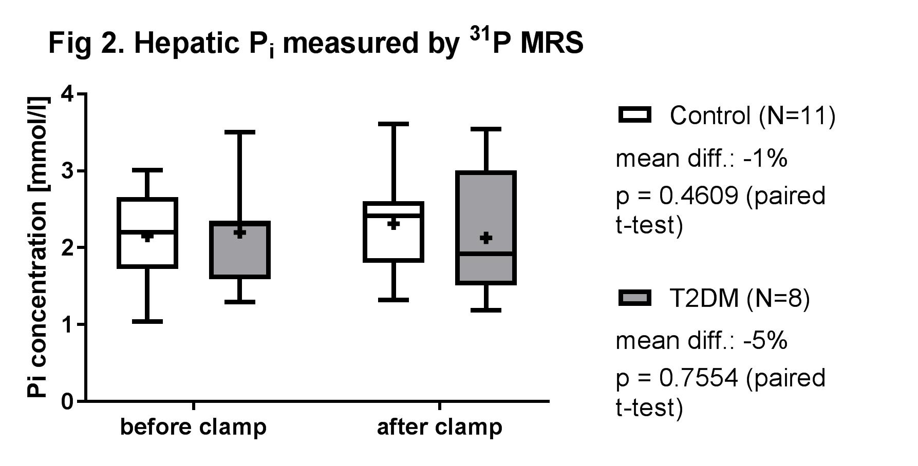
After the clamp test, hepatic γ-ATP concentrations (ATP) were significantly reduced by 18% in T2DM but not in CON (p=0.008 for T2DM vs. p=0.14 for CON, paired t-test, Fig.1). No significant change was observed in hepatic inorganic phosphate content for both groups (p=0.63 for T2DM; p=0.49 for CON, Fig. 2). At the high insulin dose, the insulin suppressed endogenous glucose production by 99.0 % and 83.6 % in CON and T2DM, respectively, indicating lower hepatic insulin sensitivity in T2DM (p=0.008) as compared to controls.DISCUSSION and CONCLUSION
This study demonstrated that the current quantitative in vivo 31P MRS allows to monitor acute changes in hepatic ATP concentrations during various metabolic conditions. During hyperinsulinemia only T2DM patients exhibited statistically significant decreases in hepatic ATP along with reduced hepatic insulin sensitivity. These data extend previous reports on lower fasting hepatic ATP levels in T2DM1.Acknowledgements
References
1. Szendroedi J, Schmid AI, Chmelik M et al. Muscle mitochondrial ATP synthesis and glucose transport/phosphorylation in type 2 diabetes. PLoS Med 2007;4(5):e154.
2. Koliaki C, Roden M. Hepatic energy metabolism in human diabetes mellitus, obesity and non-alcoholic fatty liver disease. Mol Cell Endocrinol 2013;379(1-2):35–42.
3. Weber KS, Simon MC, Strassburger K et al. Habitual Fructose Intake Relates to Insulin Sensitivity and Fatty Liver Index in Recent-Onset Type 2 Diabetes Patients and Individuals without Diabetes. Nutrients 2018;10(6):e774.
4. Laufs A, Livingstone R, Nowotny B et al. Quantitative liver 31P magnetic resonance spectroscopy at 3T on a clinical scanner. Magn Reson Med 2014;71(5):1670–1675.
5. Hamilton G, Yokoo T, Bydder M et al. In vivo characterization of the liver fat ¹H MR spectrum. NMR Biomed 2011;24(7):784–790.
Figures