1798
Dynamic contrast enhanced MRI with clinical hepatospecific MRI contrast agents in pigs: initial experience1Department of Radiology, Michigan State University, East Lansing, MI, United States, 2Department of Small Animal Clinical Sciences, Michigan State University, East Lansing, MI, United States, 3Institute of Quantitative Health Science and Engineering, Michigan State University, East Lansing, MI, United States
Synopsis
Pigs are a valuable translational biomedical tool for liver disease, and imaging can play an important role in early detection and disease characterization, but hepatic functional MRI has never been reported in pigs. Here we characterized baseline hepatic functional MRI in pigs, by performing DCE-MRI studies using two FDA-approved hepatospecific MRI contrast agents, and determined optimal animal protocols for acquiring robust data. Porcine liver has rapid accumulation of Gd-EOB-DTPA and Gd-BOPTA, following IV injection of equivalent human doses. Given the disparity in contrast agent uptake with humans for Gd-BOPTA, Gd-EOB-DTPA should be used in porcine models for biomedical imaging.
Introduction
Pigs are a valuable translational biomedical tool for liver disease, and imaging can play an important role in early detection and disease characterization, but hepatic functional MRI has never been reported in pigs. Here we characterized baseline hepatic functional MRI in pigs, by performing dynamic contrast enhanced (DCE-)MRI studies using two hepatospecific MRI contrast agents, and determined optimal animal protocols for acquiring robust data.Experimental
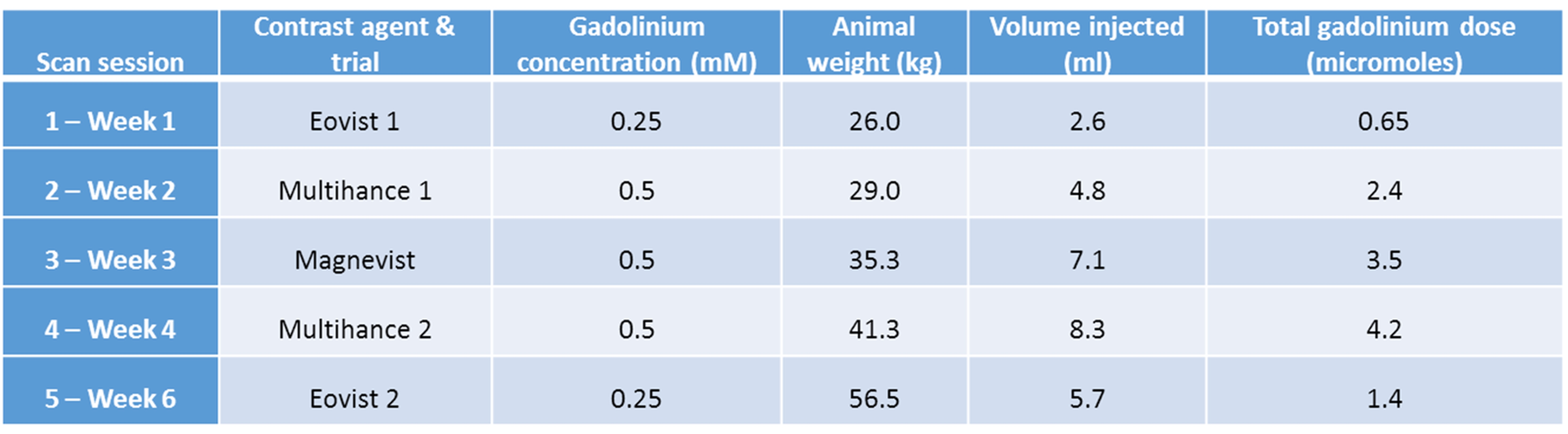
Animal experiments were approved by MSU IACUC. One 2.5 month-old, 25kg Yorkshire/PIC327 Crossbred male pig underwent 5 DCE-MRI sessions (Siemens Esprit 1.5T MRI) over 6 weeks, each time injected with a different MRI contrast agent (Table 1). Equivalent human clinical gadolinium dose was administered each session for each contrast agent, which is different depending on the agent. Experiments were staggered by 1-2 weeks.
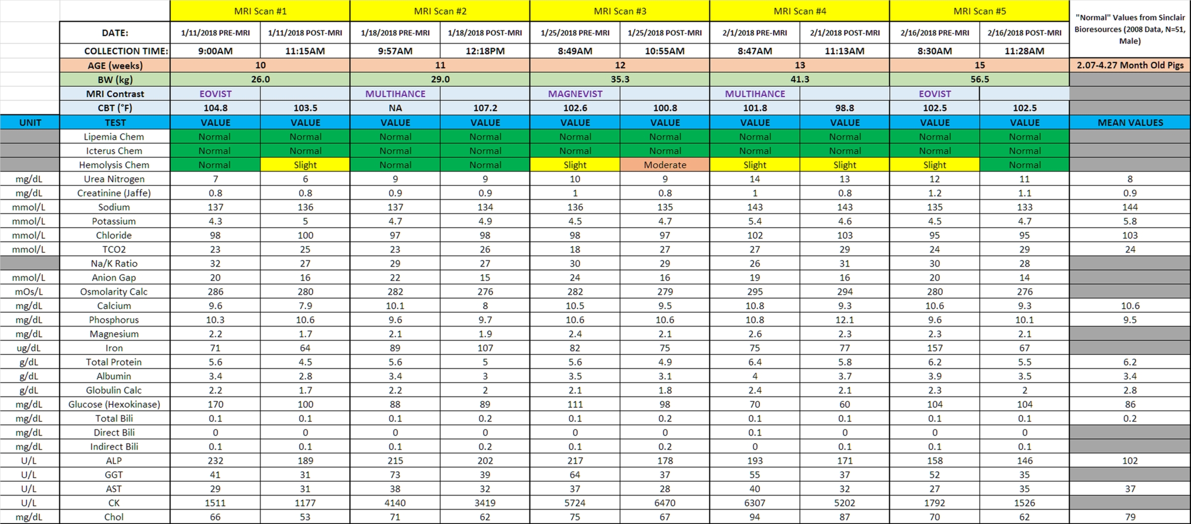
For MRI, the pig was sedated, anesthetized and intubated. Bilateral ear vein catheters were installed and blood was drawn pre- and post-MRI to measure clinical chemistry. The pig was placed inside the MRI chest-down. Anesthesia was maintained via 2-3% sevoflurane in 100% oxygen with spontaneous breathing. Vital signs were monitored, including core body temperature (CBT).
MRI protocol: DCE-MRI was performed using a T1-weighted multi-slice gradient echo VIBE sequence with Cartesian k-space filling. Scan parameters: TR=3.65ms, TE=1.77ms, FA=12°, matrix=256x256, FOV=32x32cm, slice thickness=3mm, number of slices=32, 1 frame every 13.6s.
After the fifth TR, rapid IV injection of contrast agent was performed via ear vein catheter, according to Table 1, followed by ~10mL IV bolus saline flush. Following all but the last MRI, the pig was recovered and returned to the vivarium.
Data analysis was performed in Amide 1.0.4. Cylindrical ROIs were placed over the abdominal aorta and an area of liver without major blood vessels. The same size ROI was used for all scan sessions. Percent signal enhancement was calculated from raw MRI signal intensity as (signal-baseline)/baseline. Area under the curve (AUC) was calculated to determine percent clearance by organ.
Results and Discussion
Figure 1 shows MRI before, immediately after, and 13 minutes post contrast agent injection, for Eovist, Multihance, and Magnevist. Signal enhancement in the abdominal aorta following injection is clear. Hepatic signal enhancement is seen for Eovist and Multihance, with higher enhancement for Multihance than Eovist as the dose injected was 4-fold higher.
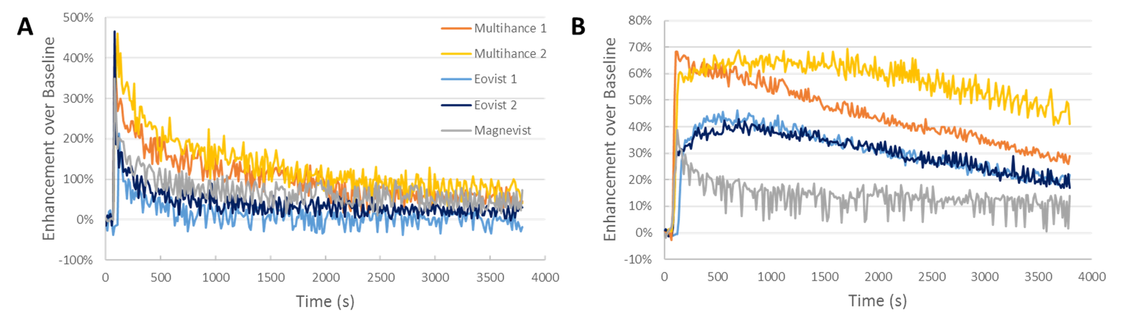
Figure 2 plots normalized signal enhancement versus time in the abdominal aorta and in the liver. Table 2 lists AUC and CBT. Both Eovist scans yielded nearly identical data, despite being performed first and last in the scan sequence and with growth of 30 kg over 5 weeks. Peak hepatic signal enhancement of 40% occurred at ~12 minutes, with gradual signal decline to 20% enhancement at 60 minutes post injection. Liver AUC and CBT were nearly identical. Distribution of Eovist occurred rapidly in the blood, on the same time scale as Magnevist.
In the first Multihance trial (Multihance 1, scan session 2, Table 1), hepatic signal enhancement was nearly instant, with a rapid, linear decrease in signal intensity for the rest of the scan session. We hypothesize that this was due to overheating; CBT after this scan session was 107.2°F. The temperature-dependence of hepatospecific MRI contrast agent influx/efflux from the liver is well known, with higher temperature resulting in faster hepatic uptake and clearance of agents (1). In the second Multihance trial (Multihance 2, scan session 3, Table 1) the addition of a cooling pad resulted in 98.8°F CBT, and MRI results showed peak hepatic signal enhancement of 65% enhancement over baseline at ~11 minutes post injection, with gradual signal decline to 45% enhancement at 60 minutes post injection. Distribution of Multihance in the blood occurred rapidly as with Eovist. ALP was slightly elevated during the entire experiment.
CK was highly elevated at scan session 2, 3 and 4 versus 1 and 5 but without control values for comparison. All other physiological measurements were normal (Table 3).
Conclusion
Porcine liver has rapid accumulation of two FDA-approved hepatospecific MRI contrast agents, Eovist and Multihance, following IV injection of equivalent human doses. For Eovist, the pig data is similar to humans, in terms of time-to-peak signal enhancement (~10 minutes) and biliary clearance rate (~50% in 1 hour). The rapid, high hepatic signal enhancement following Multihance administration differs with humans, likely due to different hepatic OATPs. There was no hepatic accumulation of the non-specific agent Magnevist. Proper animal health maintenance, especially temperature, seems essential for accurate and reproducible results. Given the disparity in contrast agent uptake with humans for Multihance, Eovist should be used in porcine models for biomedical imaging.Acknowledgements
We are grateful to: MSU CVM Radiology technicians Rex Miller and Alexis Willis-Redfern for operating the CVM MRI, MSU RATTS team for animal procedures, MSU CAR for animal care and housing, Colleen Hammond for assistance with veterinary MRI protocols and Dr. Bruno Hagenbuch, KUMC, for helpful conversations about OATPs. Grant support from the National Institutes of Health is acknowledged (R01 DK107697 to EMS).References
1. Murase K, Assanai P, Takata H, Saito S, Nishiura M. A simple and inexpensive system for controlling body temperature in small animal experiments using MRI and the effect of body temperature on the hepatic kinetics of Gd-EOB-DTPA. Magn Reson Imaging. 2013;31(10):1744-51.Figures