1787
Breathhold Black Blood Quantitative Parametric Imaging of the Liver Using Magnetization Prepared Single Shot Fast Spin Echo with DANTE Preparation1Department of Imaging and Interventional Radiology, The Chinese University of Hong Kong, Hong Kong, Hong Kong, 2Philips Healthcare, Shanhai, China, 3Philips Healthcare, Hong Kong, China, Hong Kong, Hong Kong
Synopsis
Introduction
Chronic liver disease is a major healthcare problem worldwide. Liver fibrosis is a key pathogenic and prognostic feature in most chronic liver diseases. It is reported that T1rho has a potential for detection of liver fibrosis1. However, the rich blood signal in liver can confound T1rho quantification of liver tissue. Previously, a breathhold black blood T1hro imaging sequence based on double inversion recovery (DIR) and single-shot fast spin echo (SSFSE) has been reported to address this problem2. In this work, we investigate the delay alternating with nutation for tailored excitation (DANTE)3 for this application and compare it to the previously reported methods.Methods
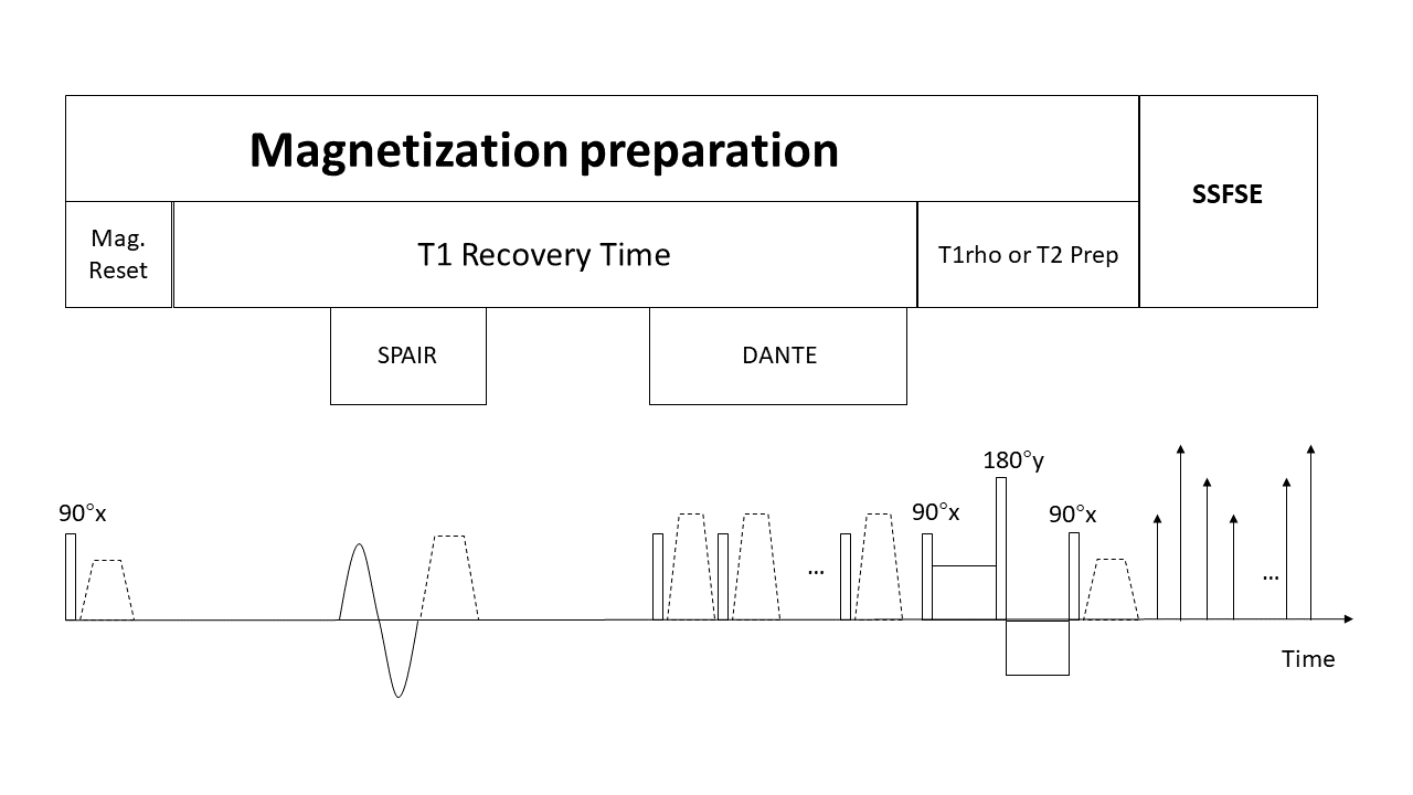
The sequence is designed so that both T1rho and T2 can be acquired in one breathhold scan with DANTE blood suppression. Figure 1 shows the sequence diagram. The DANTE preparative module contains a train of hard RF pulses interspersed with dephasing gradients. Optimized choices for this preparation module include: flip angle 12°, number of RF pulses 100, interpulse repeat time 1ms, and gradient strength 16mT/m. T1rho and T2 preparation are followed by SSFSE acquisition. The T2 preparation consists of a series of non-selective composite refocusing RF pulses 90x-180y-90x with the MLEV phase scheme. T1rho-weighted image is same as the T2-weighted image on the first time-of-spinlock (TSL) and first TE4. A spectrally selective inversion pulse was used to suppress fat signal. Acquisition parameters are: TR 2100ms, refocusing flip angle 110 °; TE for DANTE-prepared sequence is 25ms, for DIR and no blood suppression sequence 19ms; TSL for T1rho 0, 10, 30, 50ms; effective TE for T2-prep 0, 8, 17, 33ms, spin-lock frequency 500Hz; single slice; voxel size 1.5x1.5x10mm3, scan time 14.7s with a single breath-hold. Data sets were collected from a 3T Achieva MR System (Philips, Best, the Netherlands) with 32 channel cardiac coil. RF shimming and PB shimming were used to reduce B1 and B0 inhomogeneity.Results and Discussion
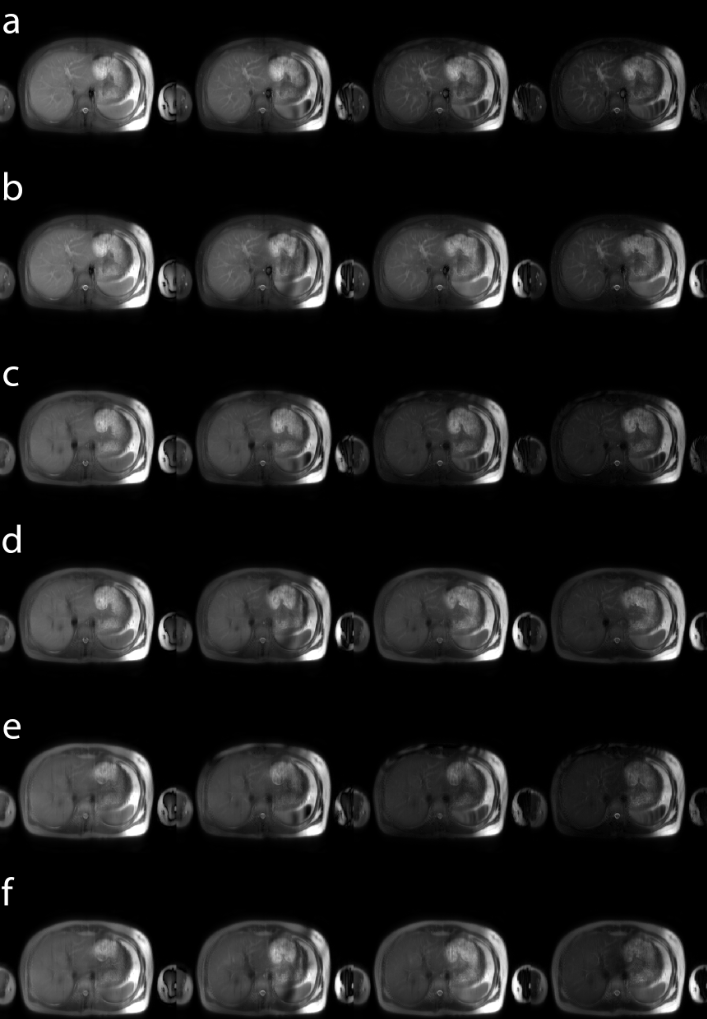
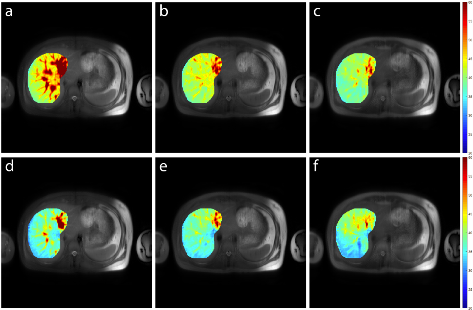
Figure 2 shows the T1rho-weighted and T2-weighted images using different black blood suppression methods. Note the proposed pulse can achieve good suppression of blood signal in the liver. Due to breathhold acquisition, all images are well registered to each other. Figure 3 shows the T1rho map and T2 map using different blood suppression methods. Note the results with SSFSE alone has elevated value of T1rho and T2 compared to the results acquired with SSFSE and additional blood suppression module. Compared to SSFSE with DIR, SSFSE with DANTE module results in a further reduced value of relaxation parameters, likely due to improved blood suppression. One possible reason causing this is that DIR may have insufficient blood suppression within a relatively thick slice (10mm) used in this study.Conclusion
We investigated a pulse sequence which combines DANTE with simultaneous T1rho and T2 acquisition in liver imaging. This pulse sequence may achieve improved blood suppression for liver relaxometry compared to the previously reported methods.Acknowledgements
This study is supported by a grant from the Hong Kong General Research Fund (GRF) 14201817 and a grant from the Research Grants Council of the Hong Kong SAR (Project SEG CUHK02).References
1. Wang YX, Yuan J, Chu ES, Go MY, Huang H, Ahuja AT, Sung JJ, Yu J. T1ρ MR imaging is sensitive to evaluate liver fibrosis: an experimental study in a rat biliary duct ligation model. Radiology. 2011 Jun;259(3):712-9.
2. Chen W, Chan Q, Wáng YX. Breath-hold black blood quantitative T1rho imaging of liver using single shot fast spin echo acquisition. Quantitative imaging in medicine and surgery. 2016 Apr;6(2):168.
3. Li, Linqing, Karla L. Miller, and Peter Jezzard. “DANTE-prepared pulse trains: A novel approach to motion-sensitized and motion-suppressed quantitative magnetic resonance imaging.” Magnetic resonance in Medicine 68.5 (2012): 1423-1438
4. Li X, Wyatt C, Rivoire J, Han E, Chen W, Schooler J, Liang F, Shet K, Souza R, Majumdar S. Simultaneous acquisition of T1ρ and T2 quantification in knee cartilage: repeatability and diurnal variation. Journal of Magnetic Resonance Imaging. 2014 May;39(5):1287-93.
Figures