1785
High-fat stimulation in healthy subjects: Longitudinal monitoring of bile acid and diffusivity changes in the gallbladder by MRS and MRI1DBMR & DIPR, University of Bern, Bern, Switzerland, 2Department of Visceral Surgery and Medicine, University Inselspital, Bern, Switzerland
Synopsis
Bile exerts multiple functions in the liver and gut and is a key player in disease processes. In this pilot study we implemented a standardized stimulation test with high-fat diet in lean physically fit individuals and performed MRS and MRI measurements longitudinally to monitor bile acid composition and diffusivity changes in the gallbladder, with the long term aim to determine a specific bile-acid-microbiota “signature”. Strongly increased bile acid and lipid resonances and reduced diffusivity after lipid ingestion were determined as well as reversal to base values within 24h, demonstrating the feasibility and potential of the method.
Introduction
Bile exerts multiple functions in the liver and is a key player in disease processes. High-fat diet is known as one of the main contributing factors for development of obesity but not each individual under high-fat intake does progress to obesity. Several lines of evidence point to bile acids (BA) and the microbiome being the culprit in pathophysiology of weight gain and associated metabolic consequences. We propose that a specific bile-acid-microbiota “signature” is present in these individuals lowering energy harvest and increasing basal energy expenditure during high-fat-diet.
The bile in the gallbladder closely reflects the bile acid pool available and being secreted into the gut. Previously it has been shown that BA can be determined non-invasively in the gallbladder by MR spectroscopy [1-3]. In a repeatability study we demonstrated stability and reliability of gallbladder spectra with relatively small coefficients of variation particularly within subjects suggesting clinical applicability of the method, especially for longitudinal studies. To our knowledge the sensitivity of the method, e.g. to determine bile composition changes upon a challenge has not been investigated.
In the current pilot study we therefore implemented a standardized stimulation test with high-fat diet in physically fit individuals and performed MRS and MRI measurements longitudinally to evaluate BA composition and diffusivity in the gallbladder, with the long term aim to determine a specific bile-acid-microbiota signature.
Subjects and Methods
Study Population: Three healthy slim male volunteers participated in this pilot study. The subjects underwent MRI examinations at three time points: at baseline after an overnight fast and 5h and 24h after starting fat ingestion (180ml, corresponding to ~800kcal Calogen liquid fat suspension). The subjects were asked to eat only moderately during the study.
All measurements were performed on a 3T-MR Scanner (Verio, Siemens) with the subjects in back position [3].
MR Spectroscopy: A single-voxel PRESS sequence with PACE-triggering (TR=1 respiration cycle, TE=35ms, 16 measurements with 4 acquisitions, 6 saturation bands, voxel size between 10x10x10mm3 and 12x12x12mm3 adapted to the individual gallbladder size) was acquired with the voxel placed in the center of the gallbladder. MRS measurement duration was ~5min, depending on breathing cycle. A non-water-suppressed spectrum was acquired for quantification [2].
MR Imaging: DWI (DWI-ZOOMit) was performed with PACE-triggering (TR=1 respiration cycle, two b-values (50, 800s/mm2), 2 repetitions, TE=46ms, slice-thickness=5mm, Matrix=90x90, FOV=200x200mm2. In addition T1 and T2 measurements were performed (not shown in detail).
Data Processing: Spectra were summed after inspection and potential discard of individual spectra. Fitting of BA and lipid peaks from the summed spectrum was performed using jMRUI AMARES [4]. The results were normalized to the non-water-suppressed spectrum. Furthermore, to avoid artificial findings due to changes of the water signal, the reciprocity principle was employed, and the metabolite values were corrected for receiver gain and transmitter value. MRI data were analyzed using in-house custom-scripts written in IDL®. For DWI regions of interest were placed manually in the center of the gallbladder and ADC determined.
Results & Discussion
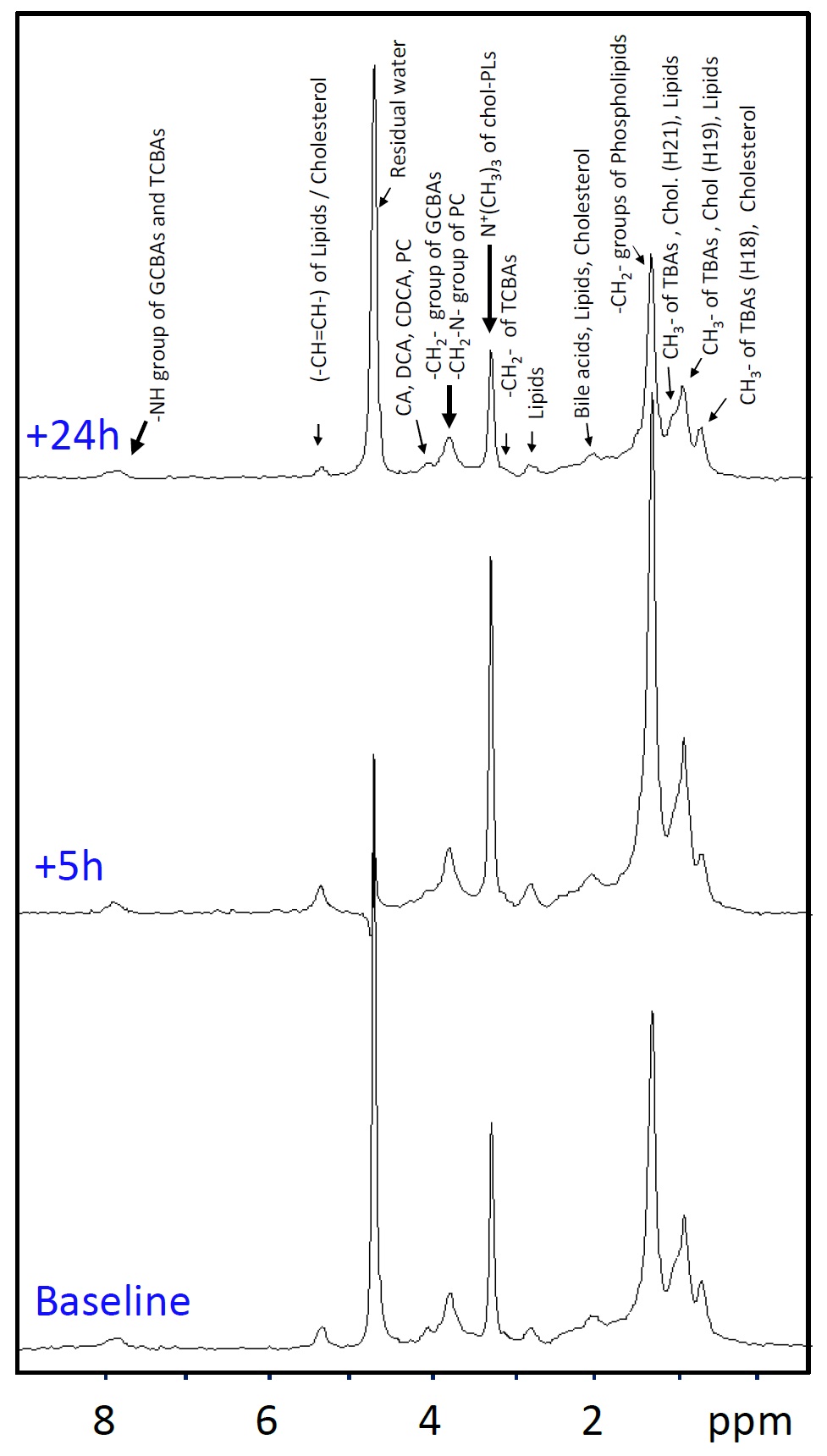
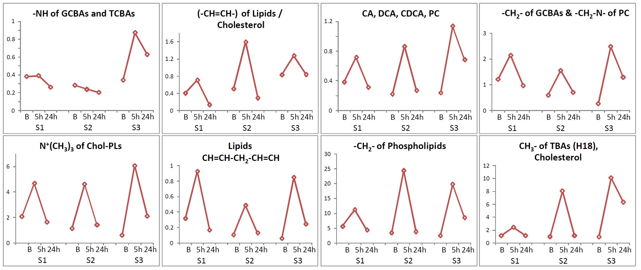
MR spectroscopy: Spectral quality was adequate in all cases, which is most likely due to measuring the volunteers in prone position with almost complete prevention of respiratory movement of the gallbladder [3]. Spectra acquired 5h after starting fat ingestion demonstrated visually a clear increase of most BA and lipid resonances compared to the spectra at baseline and a reversal after 24h (Fig.1). The results of fitting all spectra of the 3 volunteers confirmed the visual impression with an initial increase of almost all BA and lipid resonances relative to unsuppressed water after 5h and a reversal within 24h (Fig.2). Similar results were obtained when adjusting the results to receiver and transmitter gain settings, i.e. without referring to the water line These findings indicate that not only lipid resonances but also bile acids increase after a high fat diet. MR spectroscopy appears sensitive to monitor these findings.
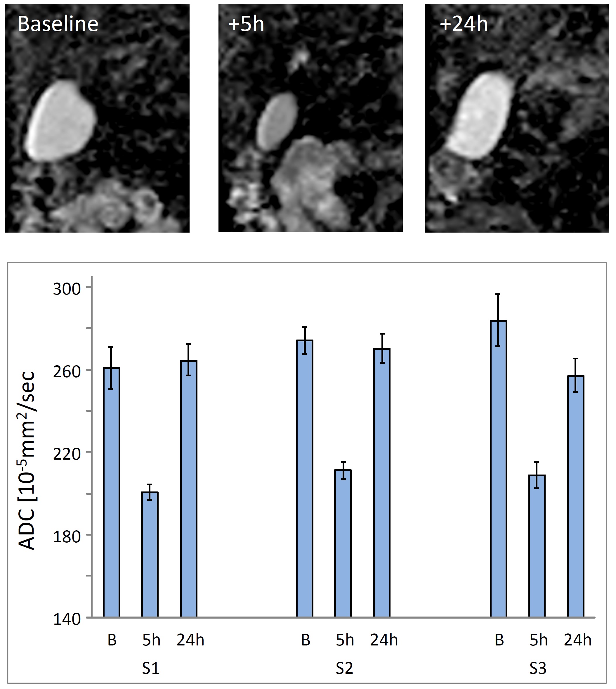
DWI: The 3-times repeated DWI measurements on three subjects before and following lipid ingestion demonstrated strongly reduced ADC values after 5h and return to baseline within 24h (Fig.3). The reduced ADC values indicate increased bile viscosity, probably due to increased lipid and BA contributions and confirmed previous finding in hamster [5]. Similar results were obtained for relaxation time measurements (results not shown in detail), with temporarily reduced T1 and T2.
Conclusion
Our study demonstrates sensitivity of MRS to monitor lipid and BA changes in the gallbladder non-invasively. These investigations build the basis for anticipated investigations testing our hypothesis of a bile-acid-microbiota “signature” in individuals, specifically adjusting energy harvest and basal energy expenditure during high-fat-diet.Acknowledgements
No acknowledgement found.References
1. Prescot
AP, Collins DJ, Leach MO, Dzik-Jurasz AS.
Human gallbladder bile: noninvasive
investigation in vivo with single-voxel 1H MR spectroscopy.
Radiology
2003;229:587-592.
2. Mohajeri
S, Ijare OB, Bezabeh T, King SB, Thomas MA, Minuk G, Lipschitz J, Kirkpatrick
I, Smith M, Smith IC.
In vivo 1H MRS of human gallbladder bile at 3 T in one
and two dimensions: detection and quantification of major biliary lipids.
NMR
Biomed 2014;27:1192-1202.
3. Diserens
G, Kreis R, Kroell D, Nett P, Stirnimann G, Vermathen P, Wiest R.
Reliable
Determination of Bile Acids from Human Gallbladder by 1H MRS - Protocol
Optimization and Estimation of Reproducibility.
Proc.Intl.Soc.Magn.Reson.Med. 2017; 25: 3549.
4. Vanhamme
L, van den Boogaart A, van Huffel S.
Improved method for accurate and efficient
quantification of MRS data with use of prior knowledge.
J Magn Reson
1997;129:35-43.
5. Tiffon
B, Parquet M, Dubrac S, Lutton C, Volk A.
In vivo gallbladder bile diffusion
coefficient measurement by diffusion-weighted echo planar imaging in hamster
fed normal and lithogenic diets.
Magn Reson Med 2000;43:854-859.
Figures