1784
Magnetic susceptibility of gallbladder stonesRakesh Kumar Gupta1, Jaladhar Neelavalli2, Manoj Kumar3, Indrajit Saha4, Pradeep Kumar Gupta5, Jitender Saini6, and Sunita Ahlawat7
1Radiology, Fortis Memorial Research Institute, Gurgaon, India, 2Philips Innovation Campus, Philips India Limited, Bengaluru, India, 3Radiology, National Institute of Mental Health And Neurosciences, BENGALURU, India, 4Philips India Limited, Gurgaon, India, 5Fortis Memorial Research Institute, Gurgaon, India, 6Radiology, National Institute of Mental Health And Neurosciences, Bengaluru, India, 7Surgery, Fortis Memorial Research Institute, Gurgaon, India
Synopsis
Quantification of volume magnetic susceptibility of extracted gallbladder stones, with 28 different types of textures, was performed using quantitative susceptibility mapping. Both dia- and para-magnetic stones are seen and the susceptibility values were found to be comparable in magnitude to those found in venous vessels and blood products – and hence could be detected in-vivo using SWI. This is in agreement with a recent work reporting visualization of gall-stones using SWI.
Introduction
Cholelithiasis and choledocolithiasis are common clinical conditions of the upper abdomen, having an incidence of 4-15% in Asia, 15% in the USA and 22% in Europe.1–3 Since its introduction 19914, magnetic resonance cholangiopancreatography (MRCP), a non-invasive imaging method, is routinely used for MRI based assessment of biliary conditions.5 However, in detecting stones smaller than 5 mm, MRCP’s sensitivity is only 62-64%.5 A recent work had pointed to susceptibility weighted imaging (SWI) as a possible an adjunct to MRCP for gall stone detection, owing to its sensitivity to magnetic susceptibility differences in SWI phase.6 Indeed ex‑vivo chemical analysis of extracted gallstones have shown that about 28% of them contain predominantly calcium salts;7 71% contain predominantly cholesterol. Even the cholesterol stones contain trace amounts of calcium salts.8 While Calcium is diamagnetic relative to water, the magnetic susceptibility of its salts is determined by the electronic structure of the compound – and hence its appearance in SWI phase. Cholesterol too, due to its lipid content, has both chemical shift and susceptibility9 (paramagnetic) difference relative to parenchyma, leading to a phase signature. However, volume magnetic susceptibility values of in-vivo gallstones is not known. As a first step towards understanding the typical magnetic properties of gall stones in the context of MR imaging - in this work we evaluated magnetic susceptibility property of gall bladder/duct extracted from patients who underwent cholecystectomy. This was done using quantitative susceptibility mapping (QSM).Materials and Methods
The study was approved by the local institutional ethics committee. A total of 32 gall stones were obtained from the surgery department of Fortis Memorial Research Institute, Gurgaon, India. After washing with distilled water, the stones were labeled and placed within two agarose gel phantoms (17 stones in phantom-1 and 15 in phantom-2) for MR imaging. One of the stones was brittle and broke down into 6 pieces, bringing the total number of individual stone samples within the phantoms to 37. MR Imaging: Both the phantoms were imaged at 1.5T Philips scanner using the SWIp sequence with parameters: TR – 52msec, TEs - 12msec, 23msec, 34msec, 45 msec, FA – 10o, BW – 493 Hz/Pix, resolution – 1 x 1 x 1 mm3. QSM maps were generated from SWIp data using the method described previously.10 Susceptibility values of individual stones were measured from the minimum or maximum intensity projection images through manual ROI selection, avoiding the edge voxels. The measurement ROI contained at least 5 voxels within the stone. The range of susceptibilities found is reported and for the stones which had similar texture, coefficient of variation (COV) measure was evaluated. It is assumed here that stones of similar texture, will have similar composition and hence comparable magnetic susceptibility values.Results
Figure - 1 shows the photographs of the stones along with their labels and the corresponding minimum (or maximum) intensity QSM maps. Of the 37 stone pieces within the phantoms, measurements could be performed in 33 stones. In 4 of them, measurement could not be performed due to significant partial voluming. Table – 1 presents the susceptibility values. Of the 32 distinct stones, 2 of them were found to be paramagnetic and the rest diamagnetic. The susceptibility values ranged from –0.102 ppm to -0.916 ppm for diamagnetic and 0.203 ppm to 0.486 ppm for paramagnetic ones. Stones having similar texture were found to have comparable susceptibility values with COV < 0.2. Stone pieces 15, 16, 17 were the pieces of the same stone and thus are expected to have comparable susceptibility value. Indeed, mean susceptibility of the three pieces was -0.488ppm with a COV of 0.057. Stones with labels 2, 11, 20 and 21 have the same texture and had mean susceptibility of 0.820 ppm and a coefficient of variation of 0.007. Similarly stones B, C, D, E, F having similar texture had mean susceptibility of -0.369 ppm and COV of 0.20; and stones K, L, M, O, with similar texture had mean susceptibility of -0.235 ppm and COV of 0.09.Discussion and Conclusion
Magnetic susceptibilities of gall-stones were quantified through ex-vivo MRI scanning using QSM. In this study, stones of 28 distinct textures were included. Stones of both positive and negative magnetic susceptibilities were observed. The susceptibility values are found to be comparable in magnitude to those of venous blood, and blood products. Thus SWI may indeed be of help (an adjunct role to play) in sensitive identification of in-vivo gall-stones.Acknowledgements
We thank Jakob Meineke and Ulrich Katscher from Philips Research, Hamburg for their support.References
1. Everhart JE, Khare M, Hill M, Maurer KR. Prevalence and ethnic differences in gallbladder disease in the United States. Gastroenterology. 1999;117(3):632-639. doi:10.1016/S0016-5085(99)70456-7 2. Aerts R, Penninckx F. The burden of gallstone disease in Europe. Aliment Pharmacol Ther. 2003;18 Suppl 3(Table 2):49-53. doi:10.1046/j.0953-0673.2003.01721.x 3. Zhang W, Jiang Z HT, R L. Epidemiology and risk factors of cholelithiasis. J Surg Concepts Pr. 2011;16(4):408-412. 4. Wallner BK, Schumacher KA, Weidenmaier W, Friedrich JM. Dilated biliary tract: evaluation with MR cholangiography with a T2-weighted contrast-enhanced fast sequence. Radiology. 1991;181(3):805-808. doi:10.1148/radiology.181.3.1947101 5. Costi R, Gnocchi A, Di Mario F, Sarli L. Diagnosis and management of choledocholithiasis in the golden age of imaging, endoscopy and laparoscopy. World J Gastroenterol. 2014;20(37):13382-13401. doi:10.3748/wjg.v20.i37.13382 6. Neelavalli J, Gupta RK, Ramaniharan AK, Gupta PK, Raj K, Bhattacharjee R. Relevance of susceptibility weighted imaging with phase (SWIp) in Cholelithiasis. In: International Society for Magnetic Resonance in Medicine. Paris; 2018. 7. Qiao T, Ma R hong, Luo X bing, Yang L qing, Luo Z liang, Zheng P ming. The Systematic Classification of Gallbladder Stones. PLoS One. 2013;8(10). doi:10.1371/journal.pone.0074887 8. Kaufman HS, Magnuson TH, Pitt HA, Frasca P, Lillemoe KD. The distribution of calcium salt precipitates in the core, periphery and shell of cholesterol, black pigment and brown pigment gallstones. Hepatology. 1994;19(5):1124-1132. doi:10.1002/hep.1840190509 9. Szczepaniak LS, Dobbins RL, Stein DT, Denis McGarry J. Bulk magnetic susceptibility effects on the assessment of intra- and extramyocellular lipids in vivo. Magn Reson Med. 2002;47(3):607-610. doi:10.1002/mrm.10086 10. Meineke J, Wenzel F, Wilkinson I, Katscher U. No significant increase of magnetic susceptibility found in subcortical gray matter of patients with Alzheimer’s Disease. In: ISMRM 25th Annual Meeting, Honolulu, HI. ; 2017:#2348.Figures
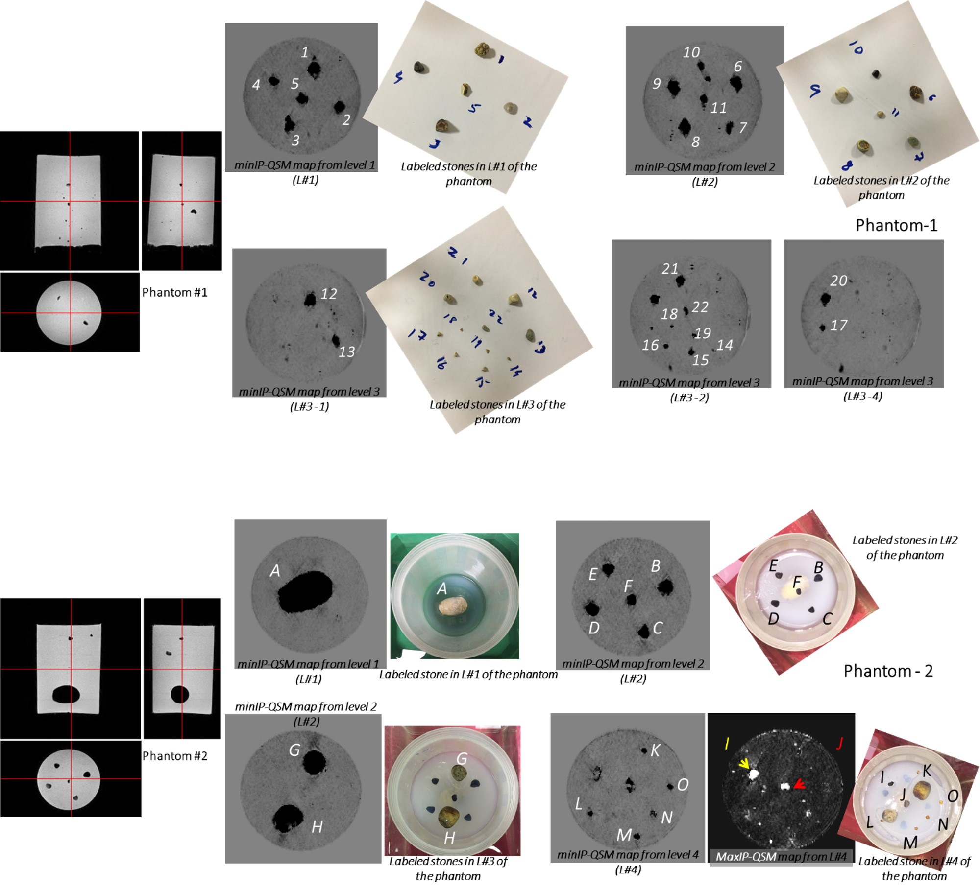
Figure – 1: Photographs of the stones imaged and the
corresponding minIP (/maxIP) QSM maps along with the corresponding labels of
the stones.
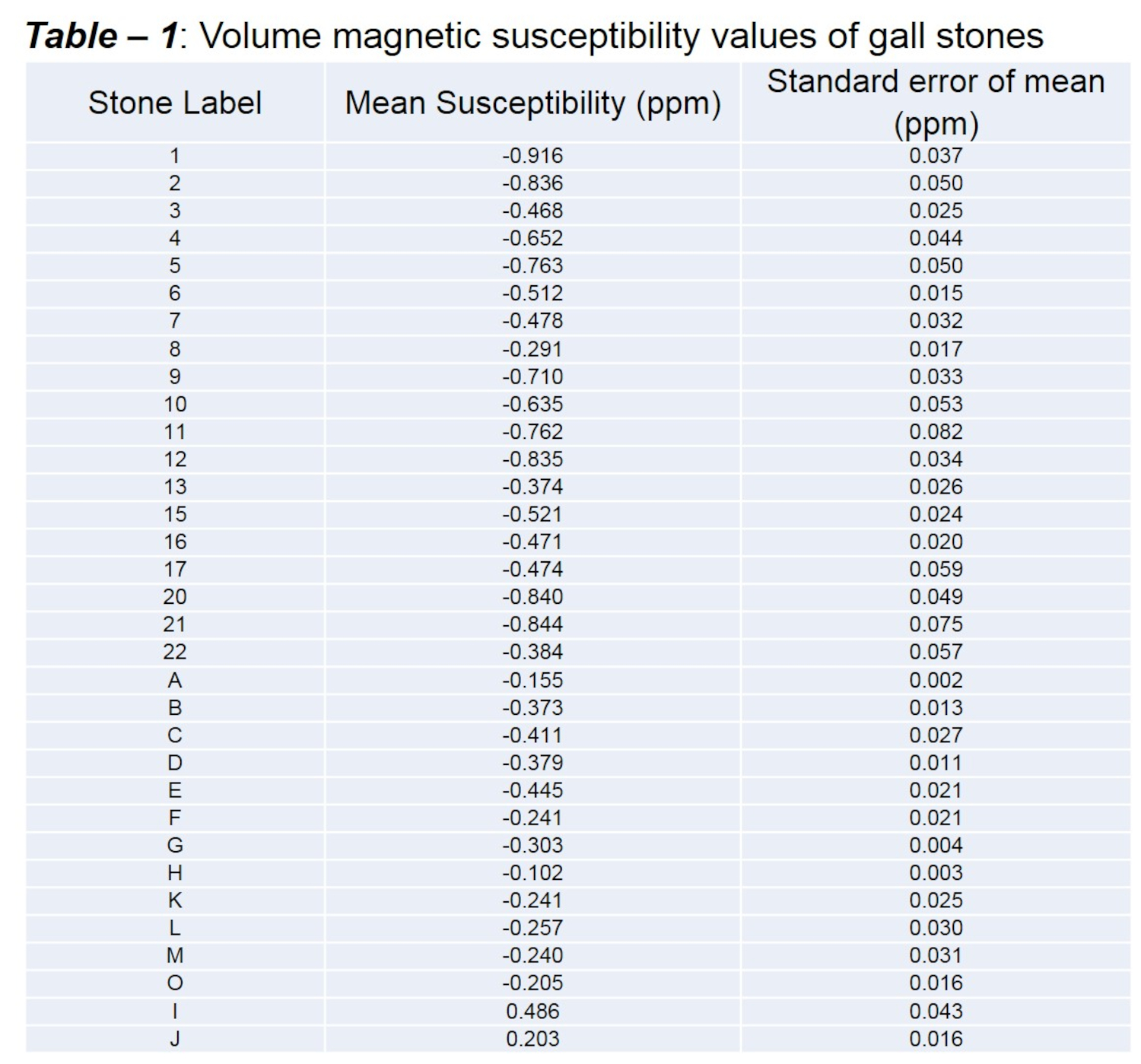
Table – 1: Volume magnetic susceptibility
values of gall stones