1757
Accuracy of multi-echo Dixon sequence in the quantification of hepatic steatosis in Chinese children and adolescents, with reference to HISTO1Shenzhen children’s hospital, Shenzhen, China, 2Siemens Healthcare, Guangzhou, China
Synopsis
This study investigated the accuracy of MRI in quantifying liver fat of 86 Chinese children and adolescents, with magnetic resonance spectroscopy (MRS) as reference. MRI and MRS were performed with multi-echo Dixon (ME Dixon) and HISTO sequence respectively to calculate hepatic proton density fat fraction (PDFF). Hepatic steatosis was diagnosed using MRS-PDFF > 5% as threshold. Spearman analysis indicated excellent correlation between ME Dixon and MRS (r>0.9,P<0.01). Bland-Altman analysis demonstrated good agreement between these two methods, indicating that ME Dixon can be an accurate way to detect hepatic steatosis in children and adolescents.
Introduction
Nonalcoholic fatty liver disease (NAFLD) is currently the outstanding cause of chronic liver disease in children and adolescents, especially in overweight and obese groups. Liver biopsy is the reference standard to diagnosis NAFLD but invasive, thus is not the best choice in clinical diagnose and follow-up. MR is widely used in clinical trials to noninvasively quantify liver fat which is suitable for children. Besides, MR can be used in early diagnosis and follow-up of NAFLD. While currently it is rarely used in Chinese children and adolescents.This study aims to investigate the accuracy of ME Dixon in quantify liver fat with magnetic resonance spectroscopy (MRS) as reference. A secondary goal was to assess the prevalence of NAFLD in overweight and obese Chinese children and adolescents.Methods
There were 86 children and adolescents enrolled in this study (mean age 13.6±1.9 years, range 9-17 years), including 65 overweight and obese children (BMI above age and gender-specific 85th/95th percentile) and 21 age- and sex-matched healthy children. They underwent MRI and MRS scan with a 3T scanner (MAGNETOM Skyra, Siemens, Healthcare, Erlangen, German). MRI and MRS were performed with multi-echo Dixon (ME Dixon) and HISTO sequence respectively to calculate hepatic proton density fat fraction (PDFF). Hepatic steatosis was diagnosed using MRS-PDFF > 5% as threshold. Spearman analysis was used to evaluate the correlation of ME Dixon and MRS. The agreement between these two methods was assessed by Bland-Altman analysis. According to the liver classification results of MRS-PDFF, sensitivity, specificity, positive predictive value (PPV) and negative predictive value (NPV) were calculated to assess the diagnostic accuracy of Dixon-PDFF.Results
The ME Dixon-PDFF in MRS ROI and the entire liver were 9.9 ± 10.3% with range 0.3% - 39.9%, and 10.6 ± 9.4 % with range 1.9% - 38.9%, respectively. The MRS-PDFF was 9.1 ± 10.0%, with range 0.5% - 37.8%. The incidence of hepatic steatosis detected by MRS-PDFF was 46.5% (40/86) of all participants, all of them belonged to overweight and obese group. Spearman analysis indicated excellent correlation between ME Dixon and MRS (r>0.9,P<0.01) (Fig 1). Bland-Altman analysis demonstrated good agreement between these two methods. With MRS-PDFF as reference, sensitivity, specificity, PPV and NPV of Dixon-PDFF were 95%, 100%, 100%, 94.9% based on ROI at the same location of MRS, and 97.5%, 88.6%, 90.7%, 96.9% covering entire liver. Four groups of typical Dixon-PDFF maps and matching MRS with varying percentages of PDFF values are presented in Fig 2.Discussion
In this prospective study of Chinese children and adolescents, 3T MRI with ME Dixon sequence accurately quantified liver fat content, with MRS (HISTO sequence) as reference. PDFF measured by ME Dixon and MRS had been validated to assess liver fat content successfully and accurately and demonstrate excellent correlation with liver biopsy (1). In our study, we observed a strong correlation between Dixon-PDFF and MRS-PDFF. Bland-Altman analysis also illustrated good agreement between these two methods. They are consistent with previous studies (2, 3), indicating that Dixon-based technique could be potentially used to quantify liver fat content for whole liver coverage. With MRS-PDFF > 5% differentiating hepatic steatosis from normal fat fraction in our study, ME Dixon achieved a good sensitivity and specificity in quantifying liver fat content. Moreover, it showed high potential in early detection of hepatic steatosis. As MRI can directly assess the entire liver fat content, thereby avoiding sampling errors when liver fat are inhomogeneous distributions. One limitation of this study is that previous study demonstrated that the best method to measure Dixon-PDFF was calculating the average value of ROI in nine Couinaud segment (4), while the Dixon-PDFF of this study was measured with ROI only at MRS location and the entire liver. A segmented measurement could be considered in the future study.Conclusion
ME Dixon shows excellent correlation and agreement with MRS in quantifying liver fat content and could be a potential tool to detect hepatic steatosis in Chinese children and adolescents.Acknowledgements
No acknowledgement found.References
1. Kang BK, Kim M, Song SY, Jun DW, Jang K. Feasibility of modified Dixon MRI techniques for hepatic fat quantification in hepatic disorders: validation with MRS and histology. The British journal of radiology. 2017:20170378.
2. Hetterich H, Bayerl C, Peters A, Heier M, Linkohr B, Meisinger C, et al. Feasibility of a three-step magnetic resonance imaging approach for the assessment of hepatic steatosis in an asymptomatic study population. Eur Radiol. 2016;26(6):1895-904.
3. Heba ER, Desai A, Zand KA, Hamilton G, Wolfson T, Schlein AN, et al. Accuracy and the effect of possible subject-based confounders of magnitude-based MRI for estimating hepatic proton density fat fraction in adults, using MR spectroscopy as reference. Journal of magnetic resonance imaging : JMRI. 2016;43(2):398-406.
4. Campo C, Hernando D, Schubert T, Bookwalter C, Pay A, Reeder S. Standardized Approach for ROI-Based Measurements of Proton Density Fat Fraction and R2* in the Liver. AJR American journal of roentgenology. 2017;209(3):592-603.
Figures

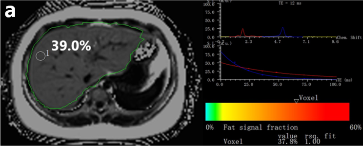
Fig.2 a


d
Examples of ME Dixon (left) and MRS (right) in four different subjects with different percentages of PDFF values. The manual measurements of ROI are shown in the ME Dixon-PDFF maps (left). The spectrum of MRS for the first acquired TE and T2 exponential decay and the estimated fat fractions (below the colored bar) are shown (right). a) to d) show different levels of fat content of 4 participants by ME Dixon (39.0%, 23.9%, 11.5%, 2.2%) and MRS (37.8%, 22.3%, 11.2%, 2.0%), respectively.