1756
Temperature sensitivities of T1, fat fraction and water resonance frequency characterised in ex vivo human livers1Oxford Centre for Clinical Magnetic Resonance Research, University of Oxford, Oxford, United Kingdom, 2Nuffield Department of Surgical Sciences, University of Oxford, Oxford, United Kingdom, 3University of Oxford, Oxford, United Kingdom, 4Department of Imaging Methods, Institute of Measurement Science, Slovak Academy of Sciences, Bratislava, Slovakia, 5Institute of Biomedical Engineering, University of Oxford, Oxford, United Kingdom, 6Wolfson Brain Imaging Centre, Department of Clinical Neurosciences, University of Cambridge, Cambridge, United Kingdom
Synopsis
MR techniques enable viability assessment of ex vivo organs for transplantation and non-invasive post-mortem examinations. However, temperature variations in ex vivo tissue and cadavers can drastically alter MR measurements of T1 and fat fraction, which risks masking underlying pathology if not considered carefully. Therefore, we investigated the changes observed in fat fraction and T1 in ex vivo human livers during a period of cooling and re-warming. Obtaining multiple measurements at different temperatures enabled determination of temperature sensitivity independent of underlying pathology, which could be used to perform a “temperature correction” of ex vivo data allowing greater sensitivity to pathological changes.
Introduction
Variations in temperature are known to affect the proton resonance frequency of water,1 the T1 of tissue2 and calculated fat fractions.3 Normally in vivo these variations are minimal and so can be ignored. However, when scanning animals under anesthesia, cadavers during post-mortems, and transplant livers ex vivo, tissue temperatures can vary by over 25°C4 and hence large changes can be expected in MR measurements. Therefore, we aimed to characterise the variation of water resonance frequency, tissue T1, and apparent fat fraction in ex vivo human livers during a period of cooling and rewarming.Methods
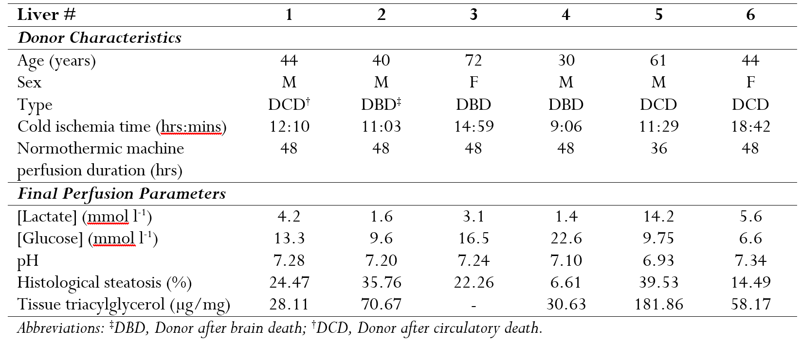
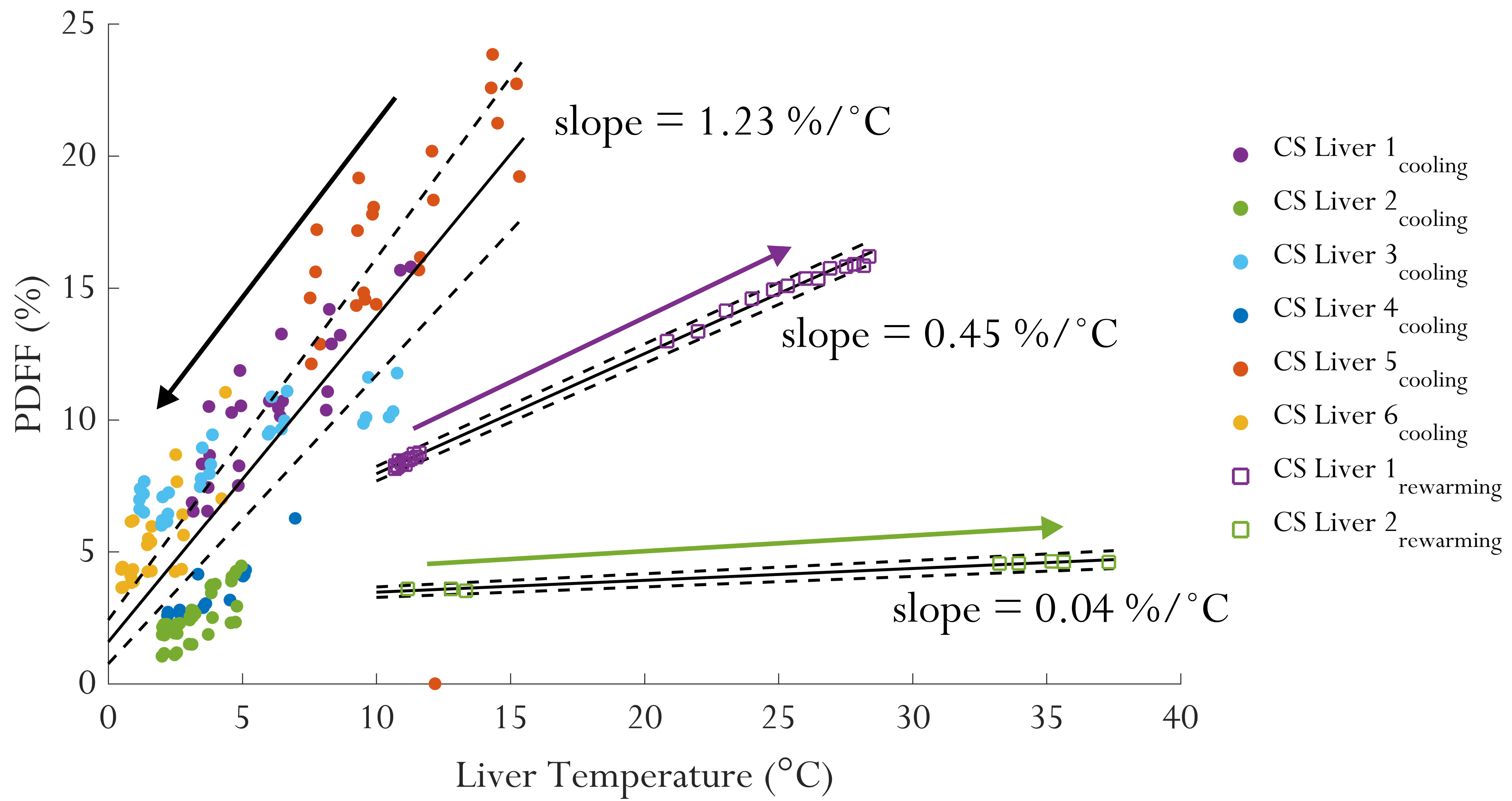
Six human livers underwent MRI examinations for 10 hrs during static cold storage. All livers were retrieved for transplantation but were declined due to visible severe steatosis. Following 48 hrs of normothermic machine perfusion on a clinical perfusion device (metra®, OrganOx, UK),5 the livers were cold flushed with 3 dm3 of histidine-tryptophan-ketoglutarate solution (Custodiol®, Essential Paramaceuticals LLC, US). Final perfusate samples were collected and the livers were placed on ice while core biopsies were taken from the right lobe. In three livers a fibre optic temperature probe (Neoptix, Canada) was inserted into the centre of the right lobe. Livers were then placed in sealed bags and transported to the MRI centre to begin scanning within 40 mins of being functional and normothermic. Following 10 hrs of cold storage two livers were placed in a water bath to recover to 12°C for one hour and then in a warmer bath to bring their temperature to ~30°C. This work has been approved by NHS Blood and Transplant (ref GP007[60]) and a national research ethics committee (ref 16/NE/0248).
T1 mapping
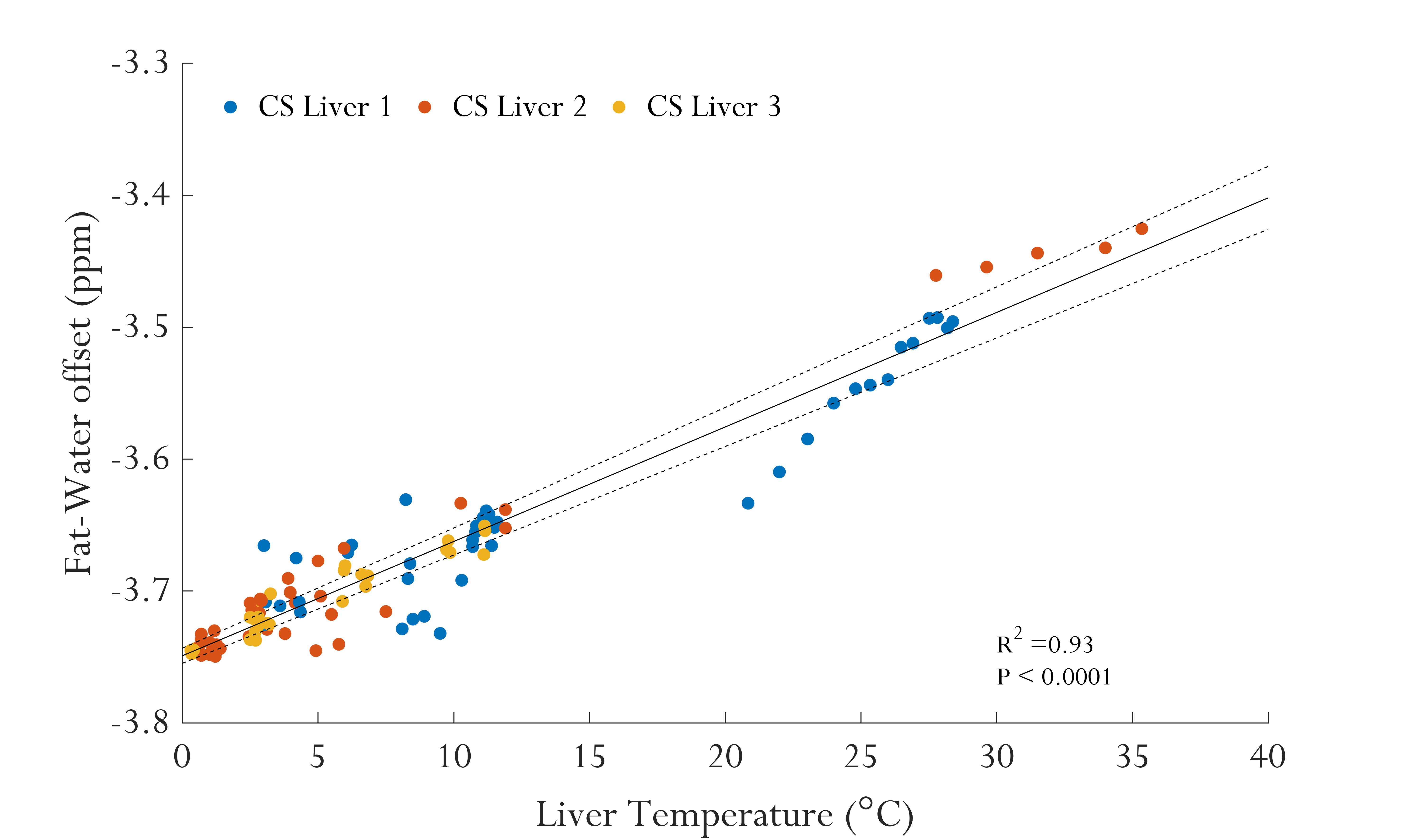
At each time point shortened modified Look-Locker inversion recovery (ShMOLLI)6 and a frequency selective variant (water only Look-Locker inversion recovery – WOLLI)7 were used to map T1 in the liver tissue. Imaging parameters were: TR/TE = 2.5/1.02 ms, flip angle = 35°, voxel size = 2.1×2.1×8 mm, 6/8 partial Fourier encoding; 2× GRAPPA, simulated R-R interval = 800 ms.
Single voxel spectroscopy (SVS)
Two SVS STEAM sequences were used to quantify the proton density fat fraction and T1 respectively. For fat fraction quantification spectra were acquired with automatically-calibrated water suppression (TR/TE = 2000/10 ms, averages = 16, measurements = 5, and voxel size = 2×2×2 cm3) and without water suppression (TR/TE = 4000, 1 average, and 3 measurements). An inversion recovery module was added to the STEAM sequence to enable a gold-standard T1 measurement using multiple inversion times (50, 500, 1218, 2385, 3553, 4750, 5500, and 7000 ms). Data analysis was completed in MATLAB, using the OXSA tool box8 for spectral fitting.
Results and Discussion
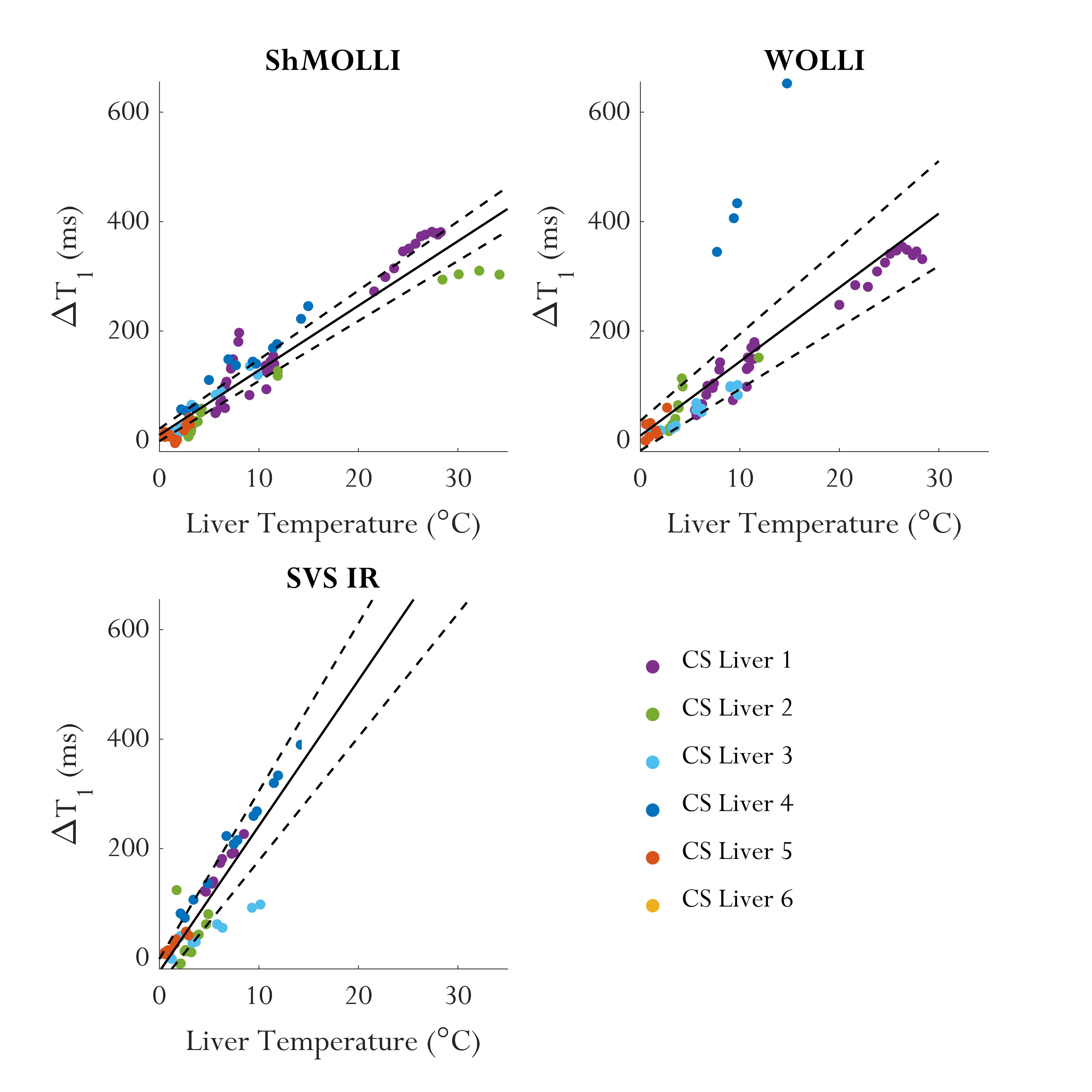
Strong linear temperature dependencies were observed in all measured parameters (P<0.0001, R2>0.67). The chemical shift difference between the (-CH2-) fat and water resonances changed at a rate of 0.0087 ppm/°C (0.0082–0.0091) in agreement with previous reports from phantom data (Figure 1).3 This was used as a surrogate marker of temperature in all livers. T1 measurements by ShMOLLI, WOLLI, and STEAM inversion recovery depended linearly on temperature as: 11.79 ms/°C (10.97–12.61), 13.52 ms/°C (11.43–15.81), and 26.03 ms/°C (22.57-30.64) respectively (Figure 2). Different temperature effects were seen between techniques due to varying sensitivities to intra/extra cellular water and fat.9 Fat fraction exhibited different temperature sensitivities during cooling, 1.23 %/°C (1.09–1.37), and re-warming, Liver 1 = 0.45 %/°C (0.44–0.46) and Liver 2 = 0.04 %/°C (0.03–0.05) as shown in Figure 3. This temperature hysteresis most likely indicates a (partial) solidification of fat at low temperatures followed by slow melting upon re-warming. All temperature dependencies are presented as mean gradient and 95% confidence intervals.Conclusion
Temperature sensitivities of T1, fat fraction and fat-water chemical shift offset were investigated in ex vivo human livers. Accurate knowledge of each technique’s temperature sensitivity in human tissue will possibly enable corrections for temperature and increase sensitivity to biological changes in temperature varying systems.Acknowledgements
This work was funded by National Institute for Health Research (NIHR) Oxford Biomedical Research Centre (BRC). LAJY is funded by the RDM Scholars programme and the MRC. CTR and LV are funded by a Sir Henry Dale Fellowship from the Wellcome Trust and the Royal Society (Grant No. 098436/Z/12/B).References
1. Kuroda, K., et al. Temperature Mapping using the water proton chemical shift: A chemical shift selective phase mapping method. Magn. Reson. Med. 38, 845-851 (1997).
2. Zech, W.-D., et al. Temperature dependence of postmortem MR quantification for soft tissue discrimination. Eur. Radiol. 25, 2381-2389 (2015).
3. Hernando, D., et al. On the confounding effect of temperature on chemical shift-encoded fat quantification. Magn. Reson. Med. 72, 464-470 (2014).
4. Levi Sandri, G.B., et al. Continuous monitoring of the liver graft temperature: relationship between bacterial contamination of the perfusion fluid and early outcome. Ann. Transl. Med. 4, 397-397 (2016).
5. Nasralla, D., et al. A randomized trial of normothermic preservation in liver transplantation. Nature 557, 50-56 (2018).
6. Piechnik, S.K., et al. Shortened Modified Look-Locker Inversion recovery (ShMOLLI) for clinical myocardial T1-mapping at 1.5 and 3 T within a 9 heartbeat breathhold. J. Cardiovasc. Magn. Reson. 12, 69 (2010).
7. Garrison, L.D., et al. Water-Only Look-Locker Inversion recovery (WOLLI) T1 mapping. in Proc. ISMRM Abstract Number: 0435 (2017).
8. Purvis, L.A.B., et al. OXSA: An open-source magnetic resonance spectroscopy analysis toolbox in MATLAB. Plos One 12, e0185356 (2017).
9. Mozes, F.E., et al. Influence of fat on liver T1 measurements using modified Look–Locker inversion recovery (MOLLI) methods at 3T. J. Magn. Reson. Imaging 44, 105-111 (2016).
Figures