1734
Liver fibrosis detection and staging in rats: a comparative study of T1 relaxation time in the rotating frame block sequence and adiabatic sequence.1Radiology department, Zhujiang hospital of southern medical university, Guangzhou, China, 2MR clinical science, Philips Healthcare, Guangzhou, China, 3Radiology department, Guangzhou, China, 4Radiology department, Shenzhen people's hospital, Shenzhen, China
Synopsis
Objective: To compare diagnostic performances on staging liver fibrosis of T1 relaxation time in the rotating frame block sequence and adiabatic sequence. Materials and Methods: 65 healthy Sprague Dawley (SD) rats were randomly divided into model and black groups. Block T1ρ and adiabatic T1ρ were performed on the rats with a 3.0-T clinical scanner. Results: T1ρ values were significantly different among stages (P <0.05), except for stages S1 and S2 with block T1ρ. AUC for block T1ρ values were 0.989, 0.924, 0.932 and 0.923, respectively. AUC for Adiabatic T1ρ values were 0.992,0.948,0.967 and 0.963, respectively. Conclusions: Adiabatic T1ρvalues had higher diagnositic performances on staging liver fibrosis in rats.
Introduction
Early diagnosis and staging of liver fibrosis are vital for clinical work1. T1 relaxation time in the rotating frame (T1ρ) have been employed to evaluate liver fibrosis stages. Recent studies, either in human or animal models, all uncovered higher mean liver T1ρ values in liver cirrhosis compared with those in normal health controls2,3,4,5. However, the relationship between adiabatic T1ρ values and fibrotic stages was rarely reported. Here we exploited the characteristics of magnetic resonance imaging (MRI) with T1ρ block sequence and adiabatic sequence in various stages of liver fibrosis, and compared their diagnostic efficiency on staging liver fibrosis and the image quality, and eventually estimated which sequence could be more potential for clinical application.Materials and Methods
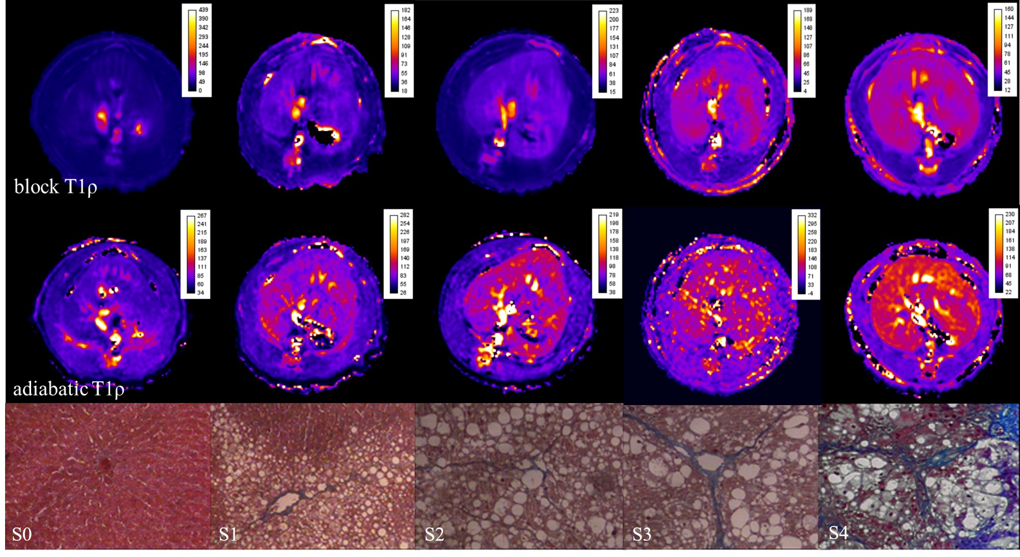
This study was approved by the institutional animal care and use committee.65 healthy Sprague Dawley (SD) rats were randomly divided into model group (50 rats) and blank group (15 rats). The hepatic fibrosis model was established by subcutaneous injection of different carbon tetrachloride (CCl4) doses. All the rats in the blank group complete T1ρ scanning one week after adaptability training. T1ρ MRI was performed on the rats with a 3.0-T clinical scanner (Achieva 3.0T TX, Philips Healthcare, Best, Netherlands. The rats were anesthetized and placed in an animal coil, with head first and in prone position. Axial scans T1WI, T2WI and T1ρ scanning were performed after 3D positioning. Main parameters of the sequences: (1) T1WI:fast spin echo sequence (TSE). TR/TE 400ms/10ms, FOV 60mm×60mm, thickness 3mm ; (2) T2WI:fast spin echo sequence (TSE), TR/TE 1080ms/120ms, FOV 60mm×60mm, thickness 3mm ; (3)Block T1ρ, fast gradient echo sequence (TFE), TR/TE 4.9ms/2.4ms, FOV 60mm×60mm, thickness 2mm , flip angle 40°. Adiabatic T1ρ, fast gradient echo sequence (TFE), TR/TE 4.2ms/2.1ms, FOV 60mm×60mm, thickness 2mm , flip angle 10°. T1ρ values were measured in workstation. A Visual score was categorized as:1:Poor,2:Fair,3:Good,4:Excellent. Liver fibrosis was staged according to the METAVIR standard. Stage F0-stage F4 of liver serial sections were stained with hematoxylin and eosin and Masson’s trichrome. Receiver operating characteristic (ROC) curve analyses were used to determine diagnostic accuracy.Results and Discussion
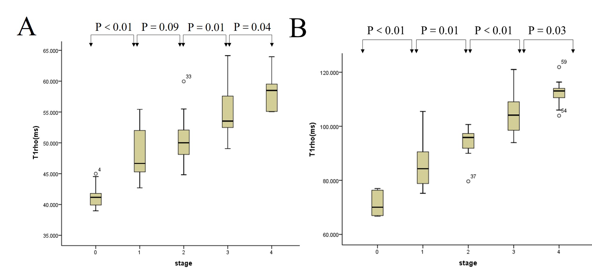
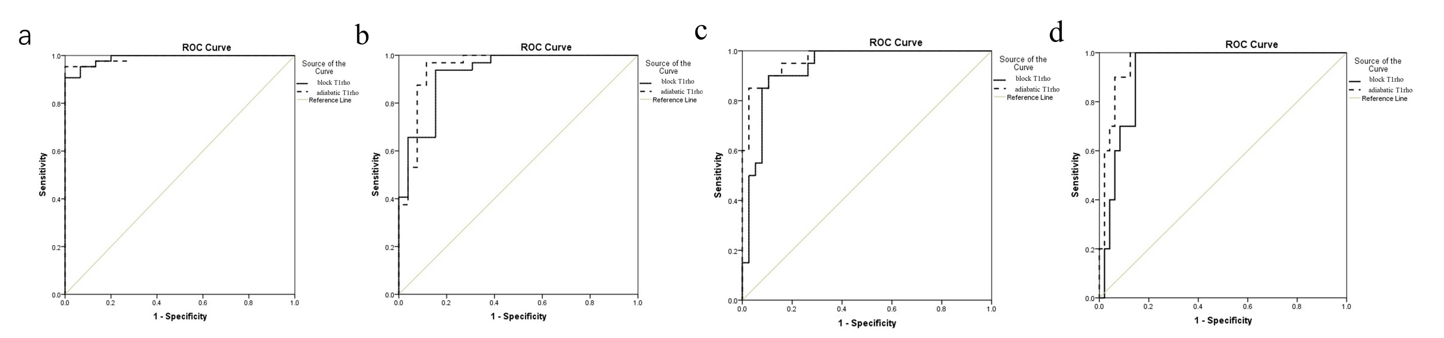
MRI and pathological examination were undertaken in 43 rats of the experimental group. S0=15, S1=11, S2=12, S3=10 and S4=10(Fig 1). Images of Adiabatic T1ρ scored significantly higher compared with routine T1ρ (P<0.05). There was a strong positive correlation between fibrosis stage and block T1ρ values (r=0.712, P< 0.001), and adiabatic T1ρ values(r=0.820, P< 0.001). One-way analysis of variance (ANOVA) showed statistical differences among different stages (P <0.05), except for stages S1 and S2 with block T1ρ (Fig 1,2). Area under ROC curve (AUC) of block T1ρ values for differentiating S0 vs S1-4, S0-1 vs S2-4,S0-2 vs S3-4 and S0-3 vs S4 were 0.989, 0.924, 0.932 and 0.923, respectively. AUC for Adiabatic T1ρ values were 0.992,0.948,0.967 and 0.963, respectively. ROC curves showed that the adiabatic T1ρ was better than block T1ρ on evaluating stages of fibrosis.Conclusions
Both T1ρ block sequence and T1ρ Adiabatic sequence had high diagnostic efficacy for the diagnosis of liver fibrosis in rats. Images of Adiabatic T1ρ had higher image quality as well. Adiabatic T1ρ values were closely associated with liver fibrosis and had higher diagnositic performances on staging liver fibrosis in rats. T1ρ adiabatic sequence may provide better clinical application than the block sequence.Acknowledgements
No acknowledgement found.References
1.Marcellin P, Gane E, Buti M, et al. Regression of cirrhosis during treatment with tenofovir disoproxil fumarate for chronic hepatitis B: a 5-year open-label follow-up study [J] . Lancet, 2013 , 381(9865) : 468-475.
2.Rauscher I, Eiber M, Ganter C, et al. Evaluation of T1rho as a potential MR biomarker for liver cirrhosis: comparison of healthy control subjects and patients with liver cirrhosis [J] . Eur Radiol, 2014 , 83 (6): 900-904.
3.Wang Y X,J Yuan, et al.T1rho MR imaging is sensitive to evaluate liver fibrosis: an experimental study in a rat biliary duct ligation model[J].Radiology,2011,259 (3):712-719.
4.Zhao F,Y X Wang,et al.MR T1rho as an imaging biomarker for monitoring liver injury progression and regression: an experimental study in rats with carbon tetrachloride intoxication[J].Eur Radiol,2012,22 (8): 1709-1716.
5. Zhang H, Yang Q, Yu T, et al. Comparison of T2, T1rho, and diffusion metrics in assessment of liver fibrosis in rats [J] . J Magn Reson Imaging, 2017 , 45(3) : 741-750.
Figures