1731
A virtual liver biopsy based on mixed MRI radiomics and biological data: a proof of conceptBenjamin Leporq1, Sophie Gaillard1, Liadeh Cuminal2, Valerie Hervieu3, Olivier Guillaud4, Jerome Dumortier4, Pierre-Jean Valette5, and Olivier Beuf1
1CREATIS CNRS UMR 5220; Inserm U1206; INSA-Lyon; UCBL Lyon 1, Université de Lyon, Villeurbanne, France, 2Department of Radiology, CHU Edouard Herriot, Lyon, France, 3Department of Pathology, CHU Edouard Herriot, Lyon, France, 4Department of Hepato-Gastro-Enterology, CHU Edouard Herriot, Lyon, France, 5Department of Radiology, CHU Lyon Sud, Lyon, France
Synopsis
Whereas NASH is associated with poor long-term outcome, widespread screening is not currently feasible given that a definitive diagnosis of NASH can only be made through liver biopsy. In this study, a virtual liver biopsy was developed with machine learning from mixed multiparametric MRI radiomics and biological data.
Introduction
In the past decade, an epidemic increase in non-alcoholic fatty liver disease (NAFLD) prevalence has been observed in Western countries and NAFLD is among the most common causes of chronic liver disease with a prevalence ranging between 17 and 46% (1). NAFLD include simple steatosis and nonalcoholic steato-hepatitis (NASH). Whereas simple steatosis has good prognosis, NASH is associated with poor long-term outcome (2) and is characterized by steatosis, hepatocyte ballooning, inflammation, with or without fibrosis at histology. About 20% of NASH patients develop cirrhosis or hepatocellular carcinoma, so that NASH has become the fastest growing cause of liver-related morbidity/mortality worldwide (3). Widespread screening is not currently feasible given that a definitive diagnosis of NASH can only be made through identification of the characteristic histopathologic pattern on liver biopsy (4). Therefore, there is a pressing unmet medical need for reliable and accurate non-invasive tools to evaluate liver steatosis, fibrosis and inflammation simultaneously. The aim of this study was to develop a virtual liver biopsy based on multiparametric MRI (mpMRI) radiomics and biological data.Methods
70 patients with chronic liver diseases, histology (ISHAK classification), blood serum markers and 3.0T mpMRI data available were retrospectively enrolled from the HEPATOMAP database. Gd-BOPTA-enhanced (t = 20 min) fat-suppressed T1w images were used as a radiomic fingerprint. PDFF-map was computed from chemical-shift-encoded acquisition according to (5). ASAT, ALAT and Gamma GT-values were recorded. From BOPTA-enhanced radiomic fingerprint, the liver were manually segmented to extract the radiome including 87 features describing shape, size, distribution and texture in images and frequency domain (Fig.1). Overall, 91 features were integrated. The learning base dimension was reduced using a backward selection by thresholding on t-test p-value (t < 0.05). Two models were established to predict advanced fibrosis (F > 2, n = 35) and inflammation (A > 1, n = 46). To predict inflammation, a k-nearest neighbor algorithm with a cosine node function and 10 neighbors was used as a classifier. To predict fibrosis, a support vector machine with a linear kernel was used as a classifier. Steatosis grades were predicted without classifier and directly from the mean PDFF-value. Internal validation was performed with a holdout cross-validation method (75% of data used for training and 25% for test).Results
Diagnosis performances to predict steatosis grades, advanced fibrosis and inflammation are summarized in Fig.2.Discussion
This work shows the feasibility to predict key liver histological characteristics (steatosis, fibrosis and inflammation) with mixed radiomics and biological data in patients with chronic liver diseases. Limitations of the study are the absence of external validation and the heterogeneity of etiologies in the database. Shared multicenter data are mandatory to extend this proof of concept to NASH only, to perform a thinner classification, and to obtain an external validation.Acknowledgements
This work was performed within the framework of LABEX PRIMES (ANR-11-LABX-0063), program "Investissements d'Avenir" (ANR-11-IDEX-0007).References
(1) Chalasani N et al. Hepatology 2012; 55(6):2005-2023. (2) Angulo P et al. Gastroenterology 2015; 149(2):389-397. (3) Satapathy SK et al. Semin Liver Dis 2015; 35(3):221-235 (4) Chalasani N et al. Hepatology. 2018; 67:328–357 (5) Leporq B et al. Eur Radiol 2013; 23(8):2175-2186Figures
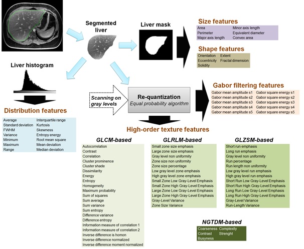
Fig.1: Radiome extraction pipeline. Size and shape features were extracted from the binary
mask. Intensity distribution features were extracted from the histogram built
with 256 bins. Images gray levels were discretized in a smaller number of gray
levels with an equal probability algorithm. Images were discretized in 8, 16,
24, 32, 40, 48 and 64 grey levels. For each discretization level, four matrixes
were built: GLCM (Gray-level co-occurrence), GLRLM (Gray-level run length), GLSZM
(Gray-level size zone) and NGTDM (Neighborhood gray tone difference) from which
characteristics were extracted, then averaged. Frequency domain-based texture
features were extracted using a Gabor filtering.
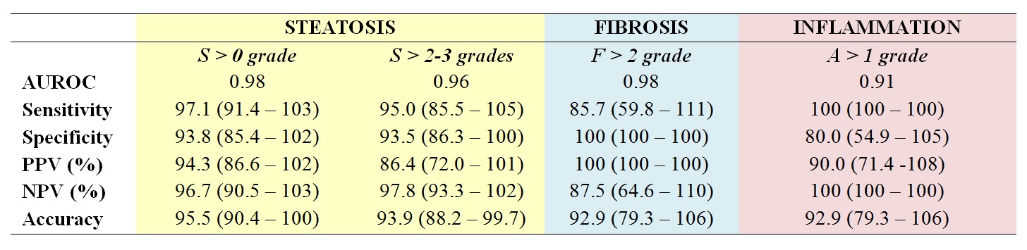
Fig.2:
Diagnosis
performances to predict key liver histological characteristics (PPV and
NPV are the negative predictive value and the positive predictive value respectively)