Dingli Ye1, Shuangyan Sun2, Jianqing Sun3, and Jihong Zhao4
1Radiology, JiLin Cancer Hospital, ChangChun, JiLin Province, China, 2JiLin Cancer Hospital, ChangChun,JiLin Province, China, 3Philips Healthcare, Shanghai, China, 4JiLin Cancer Hospital, ChangChun, JiLin Province, China
Synopsis
The purpose of this work was to use radiomics features to build a classification model that can classify breast cancer into different molecular subtypes. Our result shows that some radiomics features have great potential to be an useful index in predicting the subtype of breast cancer, therefore providing helps for the development of clinical treatment decisions for breast cancer.
Purpose
In the diagnosis and treatment of breast cancer, it is consensus for doctors that different molecular subtype requires different therapeutic schedule. 1 At present, the classification of breast cancer requires immunohistochemical examination of the lesion tissue, which is often obtained by puncture biopsy. However, for puncture biopsy, there are several disadvantages and limitations. Firstly, when multiple lesions exist in the mammary gland, the biopsy cannot obtain tissue samples of all lesions, which may affect the clinical treatment due to incomplete information acquisition. Secondly, if the operator is inexperienced or the location of the sample is not ideal, the biopsy needle cannot be inserted into and extract the right representative lesion tissue, which will finally result in wrong conclusions. Here, we investigate a nonintrusive method called Radiomics2 to classify breast cancer into different molecular subtypes. The principle of Radiomics is to extract up to thousands of texture features from medical images of tumors and build a model to predict the tumors’ properties. It has been widely applied and proven to be useful in the researches of classification and prognosis assessment of different kind of cancers .3,4 Methods
In this retrospective research, we collected 135 patients who underwent breast magnetic resonance dynamic enhancement before treatment and had confirmed postoperative pathology for breast cancer from October 2016 to September 2018. According to the definition of molecular subtype of breast cancer mentioned in the 2011 st. Gallen consensus ,5 this study include 20 cases of Luminal type A, 80 cases of Luminal type B, 18 cases of Her-2 overexpression type, and 17 cases of triple negative type. Using Bayer's Magnevis contrast agent, 15 ml of contrast agent and 15 ml of normal saline were injected intravenously at a pressure of 325psi, and the T1 hyperlipidemic images were obtained after 90-second enhancement. We use Philips Radiomics Tool to delineate the irregular region of interest (ROI) along the margin of tumor(Figure 1) and then calculate 1765 radiomics features (the algorithm is from pyradiomics6) for each patient. These radiomic features quantified tumor characteristics using tumor intensity statistics, size and shape, intensity statistics, and texture. In feature analysis and modeling stage, we used Pearson correlation, hierarchical cluster analysis and principal component analysis (PCA) to select the key features and investigated 20 classification methods for histology prediction. Multivariate models were trained on the training cohort and their performance was evaluated on the 5-fold cross-validation cohort using the area under ROC curve (AUC), accuracy, specifity and sensitivity.Result
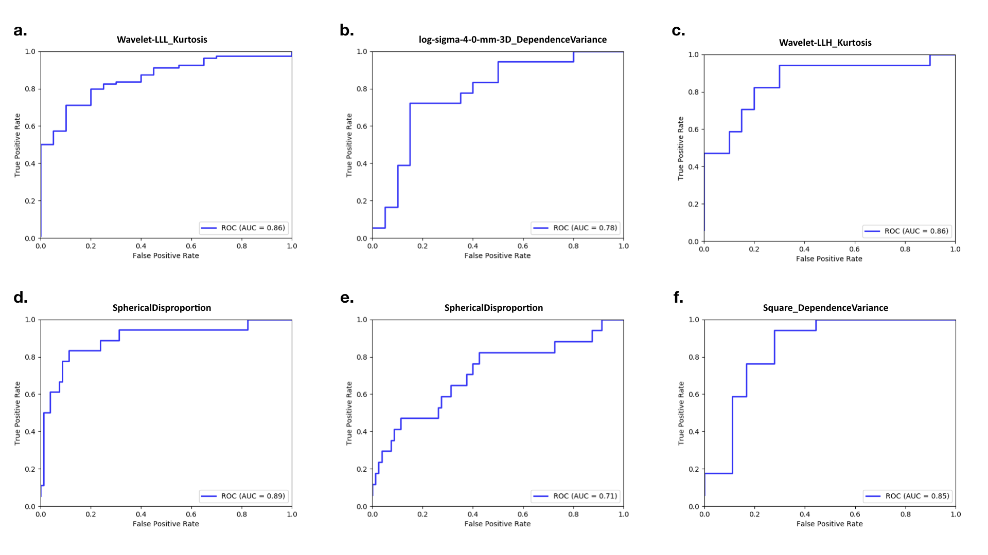
We build six models to classify 1) Luminal type A and Luminal type B, 2) Luminal type A and Her-2 overexpression type, 3) Luminal type A and triple negative type, 4) Luminal type B and Her-2 overexpression type, 5) Luminal type B and triple negative type, and 6) Her-2 overexpression type and triple negative type, respectively. The machine learning classifier of this six model are chosen to be RidgeClassifier, LogisticRegression, LogisticRegression, RidgeClassifier, RidgeClassifier, and RidgeClassifier, respectively. The performance and ROC curves of each model are shown in Figure 2 and Figure 3. The mean AUC of the six models are 0.91, 0.9, 0.97, 0.88, 0.81 and 0.89, respectively. Beside, the best ROC curves of single feature are also plotted in Figure 4, the corresponding AUCs are 0.86, 0.78, 0.86, 0.89, 0.71 and 0.85, respectively.Conclusion
Our result shows that some radiomics features have great potential to be an useful index in predicting the subtype of breast cancer, therefore providing helps for the development of clinical treatment decisions for breast cancer.Discussion
The results based on the sample size collected so far are encouraging, the results show that the texture analysis method can identify different subtypes of breast cancer, we can apply this to patients who have difficulty acquiring histopathology, provide assistance for the formulation of its clinical therapy decision. However, our research also has some deficiencies, due to the different proportion of different molecular subtypes in breast cancer patients, as a result, the number of samples varies greatly among different subtypes, and the statistical results will be biased. In the following stage, we will continue to collect related cases, increase the sample size and result credibility, so as to provide higher reliability for clinical diagnosis and treatment reference.Acknowledgements
First of all, I would like to thank my leader and colleagues for their support in my work,they are Jihong Zhao,Shuangyan Sun,Jianqing Sun,Changping Sui,Biyu Zhang,Han Zhang,Xianfeng Sun and Yijia Liu.
And secondly, I have to thank my wife Jing Ma,who is very pleasant to cook with the delicious food for me, so that I can finish my work happily.
Finally, wish the happiness be with them.
References
1.中国抗癌协会乳腺癌专业委员会. 中国抗癌协会乳腺癌诊治指南与规范(2017年版)[J]. 中国癌症杂志,2017,27(09):695-759.
2.Lambin, P., et al. (2012). "Radiomics: extracting more information from medical images using advanced feature analysis." Eur J Cancer 48(4): 441-446.
3.Aerts, H. J., et al. (2014). "Decoding tumour phenotype by noninvasive imaging using a quantitative radiomics approach." Nat Commun 5: 4006.
4.Huang, Y. Q., et al. (2016). "Development and Validation of a Radiomics Nomogram for Preoperative Prediction of Lymph Node Metastasis in Colorectal Cancer." J Clin Oncol 34(18): 2157-2164.
5.Goldhirsch A,Wood WC,Coates AS,et al.Strategies for subtypes-dealing with the diversity of breast cancer:highlights of the St Gallen International Expert Consensus on the Primary Therapy of Early Breast Cancer2011. Annals of Oncology . 2011
6.van Griethuysen, J. J. M., et al. (2017). "Computational Radiomics System to Decode the Radiographic Phenotype." Cancer Res 77(21): e104-e107.