1720
Deep Learning Assisted Fully Automatic Post-Processing for Quantitative Lung MRI1Department of Diagnostic and Interventional Radiology, University Hospital Würzburg, Würzburg, Germany
Synopsis
Functional lung MRI still suffers from a time consuming post-processing with manual image segmentation being its most time consuming part. We introduce and evaluate a deep learning based semantic image segmentation technique to enable fully automated post-processing in SENCEFUL-MRI. Obtained segmentations were compared to manual segmentations using the DICE similarity coefficient (DSC). Furthermore, quantitative ventilation values were obtained after manual and automatic segmentation. Mean DSC of the binary segmentation masks was 0.83 ± 0.09 and no significant difference in quantitative ventilation values was observed. Obtained results show that the time consuming manual post-processing in functional lung MRI can be automated by the proposed neural network.
Introduction
MRI-based
quantitative assessment of lung ventilation has been investigated recently (1–3). While techniques like Fourier
decomposition (4) or SElf-gated Non-Contrast Enhanced
FUnctional Lung imaging (SENCEFUL) (5) are promising for comprehensive
lung investigations, post-processing is still cumbersome, with manual image
segmentation being its most time consuming part.
Therefore, we now introduce
and evaluate a deep learning based semantic image segmentation technique to
enable fully automated post-processing in SENCEFUL-MRI.Methods
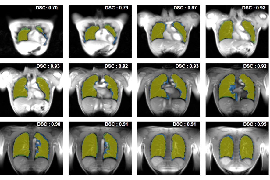
Datasets from 12 healthy volunteers were acquired using the previously proposed SENCEFUL approach (5) covering the whole lung via 10 ± 2 2D slices without gaps. Data was acquired on a 3T system (Magnetom PRISMA, Siemens Healthcare, Erlangen, Germany) applying the following parameters: TR: 2.5ms; TE: 0.7ms; flip angle: 8°; FOV: 450x450mm2; matrix size: 128x128; slice thickness: 10 mm. Until now, after registration of the images one image of the motion corrected dataset had to be segmented manually to evaluate regional quantitative ventilation (QV)(1). In the present study, this step was additionally performed by a fully convolutional artificial neural network that has been trained with 1054 single slices and according manual labels for lung segmentation from our institutional database. The network has a similar architecture as the VGG-16 network (6), however, the second part of the implemented network is represented by a decoder pattern and a final pixel classification-layer, equivalent to SegNet (7). Manual and deep learning based segmentation results were compared using the Dice similarity coefficient (DSC, (8)). Additionally, quantitative ventilation was evaluated for both manual and automatic segmentations, and compared to each other via Spearman’s rho analysis.Results
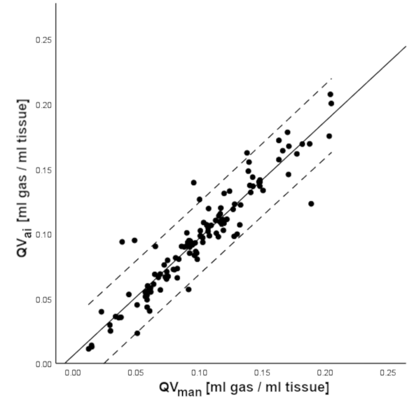
The mean DSC of the binary segmentation masks was 0.83 ± 0.09. Figure 1 presents a direct comparison of the different segmentation techniques of a representative dataset. Mean quantitative ventilation over all 2D slices was 0.10 ± 0.04 ml gas/ml lung tissue for both the manual and the deep learning approach and a significant correlation was found (rho = 0.94, p < 0.01). Figure 2 shows a scatterplot of the QV values averaged across single slices for both approaches.Discussion
Our study shows that the time consuming manual post-processing in SENCEFUL-MRI can be adequately automated by the proposed neural network. This lowers the workload of the investigator and drastically reduces the processing time without impairing quantitative ventilation results. The main metric used for evaluation of the automatic segmentation was the Dice similarity coefficient, which provided good results. Nevertheless, the pool of training data was rather small and contained labels from different operators, with varying manual segmentation style. More training data or transfer learning based on a network already trained for a similar task (e.g. semantic segmentation in cardiac MRI) would certainly further improve the overall robustness.Conclusion
Post-processing in SENCEFUL can be fully automated by introducing a convolutional neural network for semantic lung segmentation. Segmentation accuracy and the quality of further evaluations were not significantly impaired compared to time-consuming manual processing.Acknowledgements
No acknowledgement found.References
1.
Veldhoen S et al.,Radiology 2017;283:242–251.
2. Voskrebenzev A et al.,
Magn. Reson. Med.
2015;76:1542–1550.
3. Capaldi DPI et al., Radiology 2018;287:693–704.
4. Bauman G et al., Magn. Reson. Med. 2013;69:229–237.
5. Fischer A et al., NMR Biomed.
2014;27:907–17.
6. Simonyan K et al., CoRR
2014;1409.1556.
7. Badrinarayanan V et al.,
IEEE Trans. Pattern Anal. Mach. Intell.
2017;39:2481–2495.
8. Dice LR, Ecology 1945;26:297–302.
Figures