1719
Deep Learning off-resonance correction for faster free-breathing contrast-enhanced conical ultrashort echo time (UTE) MRI of the pelvis1Radiology, Stanford University, Stanford, CA, United States, 2Electrical Engineering, Stanford University, Stanford, CA, United States, 3Applied Science Laboratory, GE Healthcare, San Diego, CA, United States
Synopsis
MRI sequences with 3D cones k-space trajectories allow decreased motion artifacts while achieving ultrashort echo times (UTE). Extending readout durations allows decreased scan times but lead to worsening off-resonance artifacts. We assessed the performance of extended-readout, free-breathing UTE 3D cones MRI with and without a deep learning off-resonance correction in the evaluation of the adult pelvis. UTE imaging performed significantly better than 3D Cartesian spoiled gradient echo (SPGR) in noise, and after off-resonance correction also performed significantly better in artifact reduction.
Introduction
3D pelvic MRI can be critical for the accurate detection and staging of anorectal, cervical, and endometrial malignancy as well as characterizing complex pelvic anatomy and pathologies including anorectal fistulas1,2. Current 3D Cartesian trajectories often require five minutes or more of scan time with resulting motion artifacts. 3D cones trajectories are more scan-time efficient and motion robust than 3D Cartesian trajectories3,4. However, with 3D cones UTE, there is flexibility for a tradeoff between scan-time efficiency and off-resonance artifacts, which manifest as blurring. Previous studies have demonstrated that machine learning is capable of correcting for off-resonance (OR) artifacts, enabling scan time reduction while maintaining image quality5.
The goals of the present study were (1) to demonstrate feasibility of 3D cones UTE imaging in the pelvis and (2) to assess artifact reduction using machine learning based OR correction.
Methods
This was a prospective study with IRB approval and informed consent. We compared the 3D cones UTE trajectory (FOV 44 cm, 416 x 416 reconstruction matrix, ST 1.8 mm, SS 0.9 mm; Figure 1) to a standard free breathing 3D spoiled gradient echo (SPGR) sequence (FOV 44 cm, 512 x 512 matrix, sample TR 4.7, sample TE 1.1, ST 1.4 mm, SS 0.7 mm) with gadobutrol (Gadavist, Bayer, Whippany, NJ; 0.1 mL/kg). The base readout duration was 1.1 ms and readout duration was initially extended by a factor of 3, 5, 7, and 9 to qualitatively assess the severity of blurring and ensure high-quality diagnostic images even prior to off-resonance correction (Figure 2) .
OR correction was performed with a Deep Learning model, Off-ResNet (Figure 1). Inclusion criteria were adult age and referral for 3T contrast-enhanced pelvic imaging. Exclusion criteria were musculoskeletal and prostatic indications. 21 sequential patients were included, 11 female, mean age 54 (34-81) years.
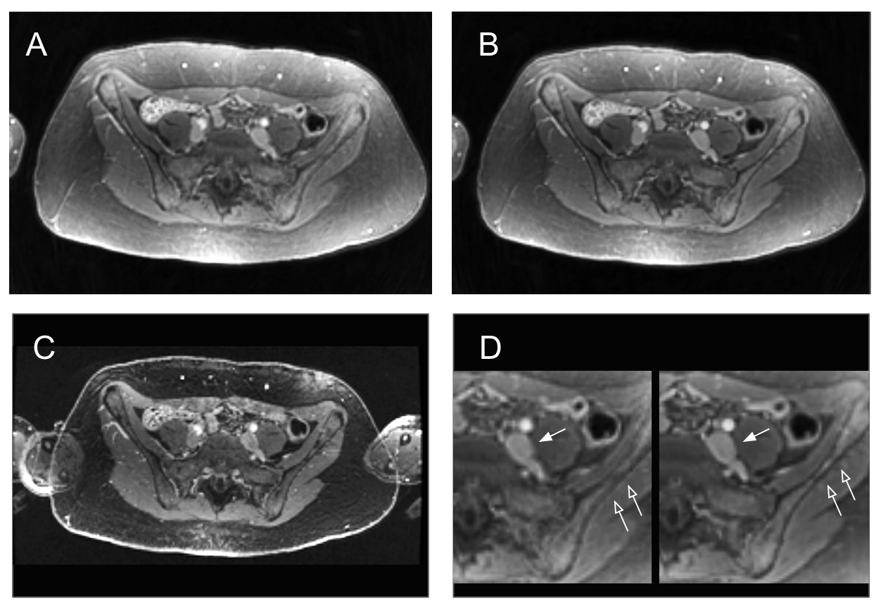
Based on variable levels of scanning dependent on clinical indication, multiple anatomic levels were chosen based on reproducibility and selected by highest level available for both subject and case control from (1) mid-sacroiliac joint, (2), acetabular roof, (3) mid-femoral head. Uncorrected and OR-corrected UTE images were presented in comparison to each other and separately presented in comparison to SPGR, with screen position randomized. Representative images are presented in Figure 3. Images were compared according to predetermined criteria judging artifacts, anatomic visualization, noise, contrast, and sharpness (Table 1). Two readers not involved in the selection of the stretch factor (PS and VS, both with five years experience interpreting body MRI) independently scored the 63 sets of images in a blinded, randomized fashion.
Inter-reader agreement was assessed using the weighted Cohen’s kappa statistic. Image quality scores between pulse sequences were assessed using the two-tailed Wilcoxon signed rank test. Scored parameters that had fewer than six effective samples were not evaluated for significance. P<0.05 following Bonferroni correction for multiple comparisons was considered significant.
Results
Based upon the initial results (Figure 2), a readout extension factor of 5 was selected (readout duration 5.5 ms, for a sample scan time 1:53 vs 6:11 without readout extension and 5:04 for SPGR). In the 21 patients, clinical indications included perianal fistula (n=5), anorectal malignancy (n=5), urinary tract malignancy (n=1), cervical/endometrial malignancy (n=5), benign uterine pathology (n=2) and scrotal pathology (n=2).
The weighted Cohen’s kappa statistic for all observations was 0.51 (95%CI 0.23-0.37), indicating fair agreement between the two readers6.
Uncorrected UTE performed significantly better than SPGR better for perceived image noise (p=0.0001), while OR-corrected UTE performed significantly better for both perceived noise (p<0.0001) and image artifacts (p<0.005). Remaining comparisons were nonsignificant after Bonferroni correction for multiple comparisons.
Discussion
Both OR-corrected and uncorrected UTE were noninferior to the existing SPGR protocol, and in fact were significantly less noisy. Following OR-correction, the reduction in image artifacts was also significant. This is in keeping with previous results showing superior perceived SNR and fewer artifacts in pediatric abdominal imaging4.
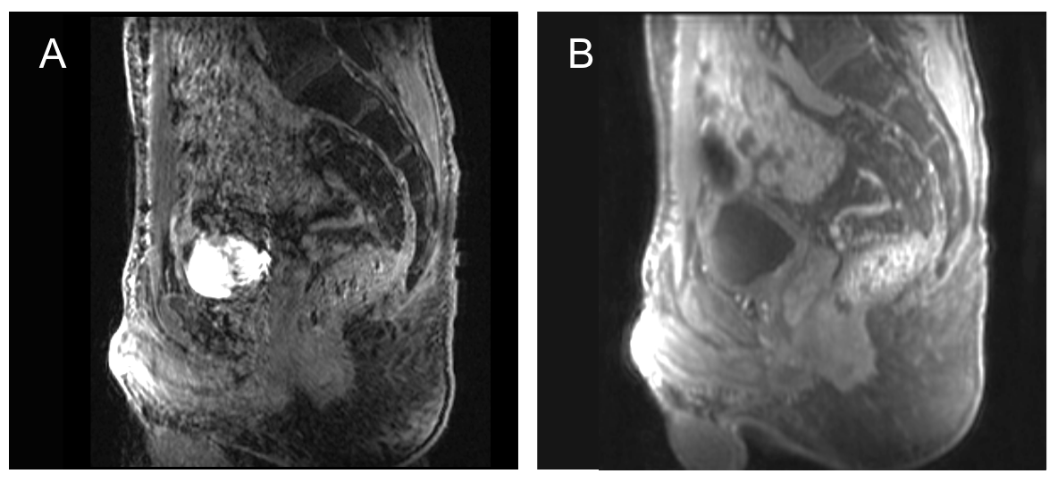
The overall performance of conical UTE in the axial plane is particularly significant given the additional advantages over Cartesian imaging in multiplanar reconstruction and imaging time (Figure 4). While nonsignificant, there was a trend towards improvement in image sharpness when OR-corrected UTE images were compared with uncorrected images (p=0.025) even without maximizing readout duration. Clinical implications of image sharpness are significant and these findings emphasize the contribution of Deep Learning off-resonance correction.
This study was limited by a small sample size and lack of quantitative image quality assessment features. Given promising initial results, further development and performance testing are warranted.
Conclusion
Conical UTE imaging in the pelvis is feasible given noninferior to superior performance across multiple qualitative measures when compared to Cartesian SPGR imaging. OR-correction with a machine learning algorithm improves image quality while decreasing scan time and merits further development.Acknowledgements
Grant support of NIH R01EB009690, R01 EB026136, R01HL136965, and GE Healthcare.References
1. Jentzsch T, Vlachopoulos L, Fürnstahl P, Müller DA, Fuchs B. Tumor resection at the pelvis using three-dimensional planning and patient-specific instruments: a case series. World J Surg Oncol. 2016 Sep 21;14(1):249.
2. Imaging and Surgical Management of Anorectal Vaginal Fistulas. VanBuren WM, Lightner AL, Kim ST, Sheedy SP, Woolever MC, Menias CO, Fletcher JG. Radiographics. 2018 Sep-Oct;38(5):1385-1401.
3. Zucker EJ, Cheng JY, Haldipur A, Carl M, Vasanawala SS. Free-breathing pediatric chest MRI: Performance of self-navigated golden-angle ordered conical ultrashort echo time acquisition. J Magn Reson Imaging. 2018 Jan;47(1):200-209.
4. Roh AT, Xiao Z, Cheng JY, Vasanawala SS, Loening AM. Conical ultrashort echo time (UTE) MRI in the evaluation of pediatric acute appendicitis. Abdom Radiol.
5. Zeng DY, Shaikh J, Nishimura DG, Vasanawala SS, Cheng JY. Deep Residual Network for Off-Resonance Artifact Correction with Application to Pediatric Body Magnetic Resonance Angiography with 3D Cones. arXiv:1810.00072, 2018.
6. Landis JR, Koch GG. The measurement of observer agreement for categorical data. Biometrics. 1977;33:159–174.
Figures

Figure 1.
A) An RF-spoiled GRE sequence with UTE is designed with a 3D cones k-space sampling trajectory. The data collection of each cone interleaf is ordered using the golden-ratio increment.
B) Network architecture for Off-ResNet, a pre-trained three-layer residual network for off-resonance artifact correction.
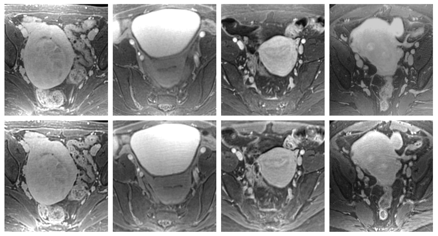
Figure 1.
Row 1: Images through the level of the uterine body in four patients undergoing evaluation for uterine fibroids without readout extension.
Row 2: Corresponding images in the same four patients when readout factors were extended (left to right) by factors of 3, 5, 7 and 9. Representative imaging times were 6:11 (no readout extension), 2:29 (readout extension factor = 3), 1:53 (readout extension factor = 5), 1:34 (readout extension factor = 7), and 1:29 (readout extension factor = 9).