1715
Fully Automated Segmentation of Cervical Cancer in Diffusion MR Imaging Using U-Net Convolutional Neural Networks1Dept. Medical Imaging and Intervention, Chang Gung Memorial Hospital, Linkou, Taiwan, 2Dept. Medical Imaging and Radiological Sciences, Chang Gung University, Taoyuan, Taiwan
Synopsis
The aim of the study is to evaluate the performance of U-Net in tumor segmentation on diffusion MR imaging for patients with cervical cancer. Diffusion weighted imaging of b0, b1000 and ADC maps were used for training. The ADC histogram parameters of predicted region of tumor were assessed for accuracy and reproducibility. The results show the triple-channel training algorithm exhibited the best performance in both training and testing datasets. The predicted voxels of tumor can be used to generate the volumetric ADC data for Radiomics study.
Purpose
Cervical cancer ranks the fourth for both incidence and mortality in the world. Diffusion weighted (DW) MR imaging demonstrated potentials in integrating ADC values with clinical information to improve the predictive accuracy 1. MRI is the best for assessing tumor border and local issues and provide the foundation of volumetric quantitative measurement. The aim of this study is to evaluate the performance of U-Net in tumor segmentation on diffusion MR imaging in patients with cervical cancer.METHODS
Among the 169 patient databank, 144 patients were used for the training phase, while another independent 25 patients were used for the testing. The sagittal DW imaging of b0, b1000 and ADC maps of each slice of each patients were used as the input for the subsequent training.
The end-to-end neural network was adopted for tumor segmentation based on the U-Net architecture using a fully convolutional network 2. The architecture combined a contracting (down-sampling) path to capture context and a symmetric expending (up-sampling) path that enables precise localization 3. A total of 17 convolutional layers were employed in the network. Image augmentation was performed with cropping, rotation, and shifting, which generated 61320 images for each data sets. Seven combinations from the 3 image sets (b0, b1000 and ADC) were used as the input sources in different channels for training.
To test the reproducibility of the training model, a repeat training procedure was performed using the identical parameters. After building the models, we examined the accuracy of the models using the testing dataset images. The accuracy of segmentation was evaluated using Dice similarity coefficient (DSC),. The histogram parameters of ADC values (mean, minimum, 10th, 25th, 50th, 75th, 90th percentiles, maximum, kurtosis, skewness and standard deviation) extracted by the predicted algorithm was correlated with those from manually labeled using Pearson correlation. The reproducibility of the training was assessed using the Intraclass Correlation Coefficient (ICC) between the ADC histogram parameters of ROIs extracted from the 1st and 2nd training algorithms.
RESULTS
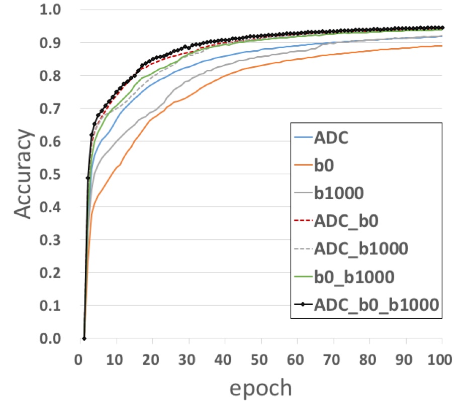
Performance in Training phase. In the training stage, The use of triple channel input (ADC+b0+b1000) exhibited the fastest learning efficacy to reach the plateau with the highest accuracy of 0.946. The use of single channel of b0 had the lowest learning efficacy with the plateau accuracy of 0.891 (Fig1).
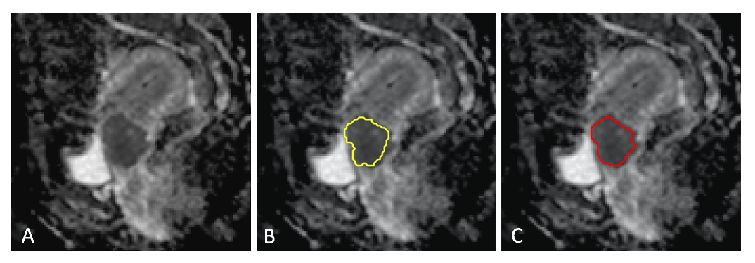
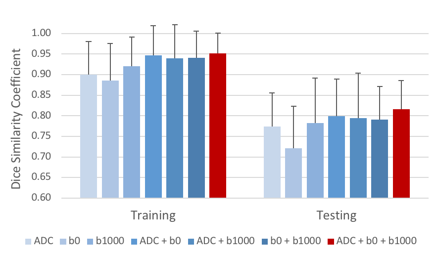
Performance of in validation phase. Fig.2 illustrates an example of fully automated tumor segmentation for a patient with cervical cancer in the testing dataset. The predicted ROI, which was generated using the triple-channel training algorithm (ADC + b0 + b1000), was segmented correctly at the corresponding tumor location as in the labeled image. Fig.3 plots the accuracy of segmentation for various combinations of source images that fed to training, the triple channel model exhibited the highest DSC (0.95±0.05 and 0.82±0.07 respectively for the training and testing datasets).
Tumor ADC Histogram parameters. All the ADC histogram parameters of the tumor were highly correlated between the labeled and predicted ROIs. (r = 0.25 – 0.9, p <0.001 for all). The reproducibility of the histogram parameters between the 1st and 2nd trainings were high for all the histogram parameters (ICC = 0.79 – 0.98).
DISCUSSION
The current study shows that combining b0, b1000 and ADC images as 3-channels input exhibits the best performance among various combinations of datasets in both the training and testing phases. Previous report showed that the ADC histogram extracted from pretreatment MRI improves the prediction for overall survival and disease-free survival for patients with stage IB-IV cervical cancer following chemoradiotherapy 1. The current study demonstrate that the predicted ADC histogram parameters of tumor are highly reproducible and correlated well with those from manual ROI. This implies that the algorithm based on U-Net architecture may boost the extraction of radiomics features by automatic contouring of the tumor in cervical cancer, which could help oncologists accentuate the follow-up for patients to assess for recurrence.CONCLUSION
U-Net based convolutional neural networks can perform accurate localization and segmentation of cervical cancer on diffusion MR imaging. The predicted voxels of tumor can thus be used to generate the volumetric ADC data that may be used as biomarkers for Radiomics study.Acknowledgements
Funding: Supported by National Science Council (Taiwan) MOST 106-2314-B-182A-016 -MY2References
- Lin G, Yang LY, Lin YC, et al. Prognostic model based on magnetic resonance imaging, whole-tumour apparent diffusion coefficient values and HPV genotyping for stage IB-IV cervical cancer patients following chemoradiotherapy. Eur Radiol. 2018.
- Shelhamer E, Long J, Darrell T. Fully Convolutional Networks for Semantic Segmentation. IEEE Trans Pattern Anal Mach Intell. 2017;39(4):640-651.
- Ronneberger O, Fischer P, Brox T. U-Net: Convolutional Networks for Biomedical Image Segmentation. 2015; Cham.
Figures