1709
Partial velocity-compensated optimized diffusion encoding for combined motion compensation and residual vessel signal suppression in liver ADC mapping1Technical University of Munich, Munich, Germany, 2Philips Healthcare, Best, Netherlands, 3Philips Healthcare, Tokyo, Japan, 4Philips Healthcare, Hamburg, Germany
Synopsis
Despite its strong clinical significance in lesion detection and tumor staging, liver DWI remains challenged by its strong sensitivity to motion effects. Motion-compensated diffusion encoding schemes have recently been proposed to improve DW liver signal homogeneity especially in the left liver lobe, a region typically affected by cardiac motion. However, motion-compensated diffusion encoding is associated with hyperintense vessel signal even at high b-values, which can obscure lesion detection. The present work proposes a partial velocity-compensated diffusion encoding using asymmetric diffusion gradients for combined motion compensation and residual vessel signal suppression in liver DWI, optimized for short echo times.
Purpose
Diffusion-weighted imaging (DWI) remains a valuable tool in liver lesion detection and there is an ongoing interest in ADC mapping for tumor staging and therapy monitoring1,2. However, cardiac motion remains a major source of signal intravoxel dephasing and ADC overestimation in the left liver lobe. Motion-compensated diffusion encoding designs have recently been proposed as alternatives in overcoming the low efficiency of cardiac triggering in removing the effect of cardiac motion from liver DWI3,4,5. However, when motion-compensated diffusion encoding is used, vessels with predominantly flow (coherent) motion might remain hyperintense even at high b-values, resulting in high ADC values. Partial velocity-compensated diffusion encoding, combining velocity compensation using symmetric bipolar gradients in the frequency and slice encoding axes and traditional Stejskal-Tanner diffusion gradients in the phase encoding axis, has recently been shown to enable reduced motion sensitivity in liver DWI while maintaining good vessel signal suppression6. Optimized asymmetric velocity-compensated diffusion gradients have also been recently proposed to further reduce the echo time7,8. The present work proposes a novel method combining partial velocity-compensated diffusion encoding with optimized asymmetric velocity-compensated diffusion gradients and investigates the variation between the ADC values reported by different diffusion encoding waveforms.Methods
Partial optimized asymmetric velocity-compensated diffusion encoding scheme (asym vc-pgsep)
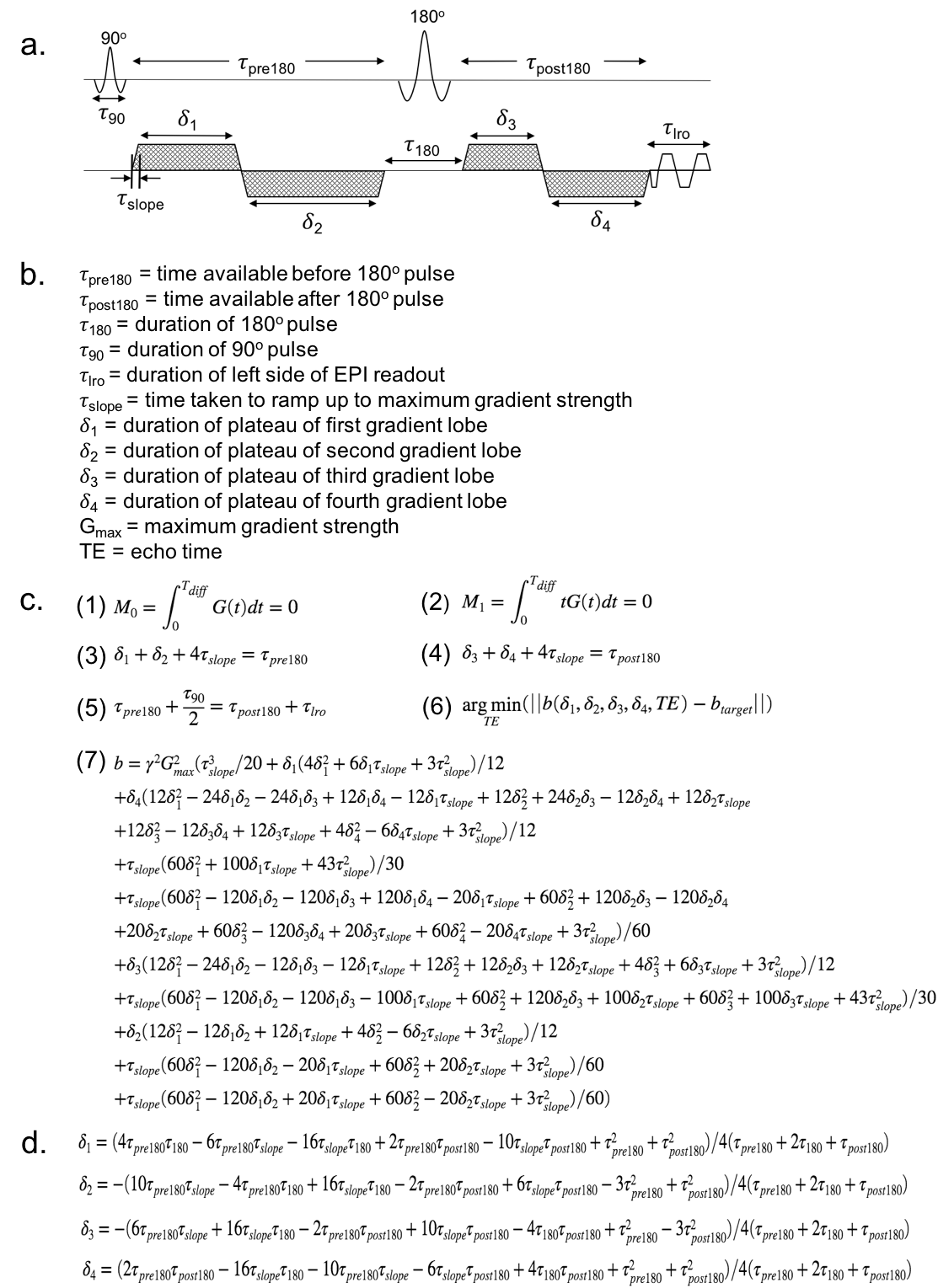
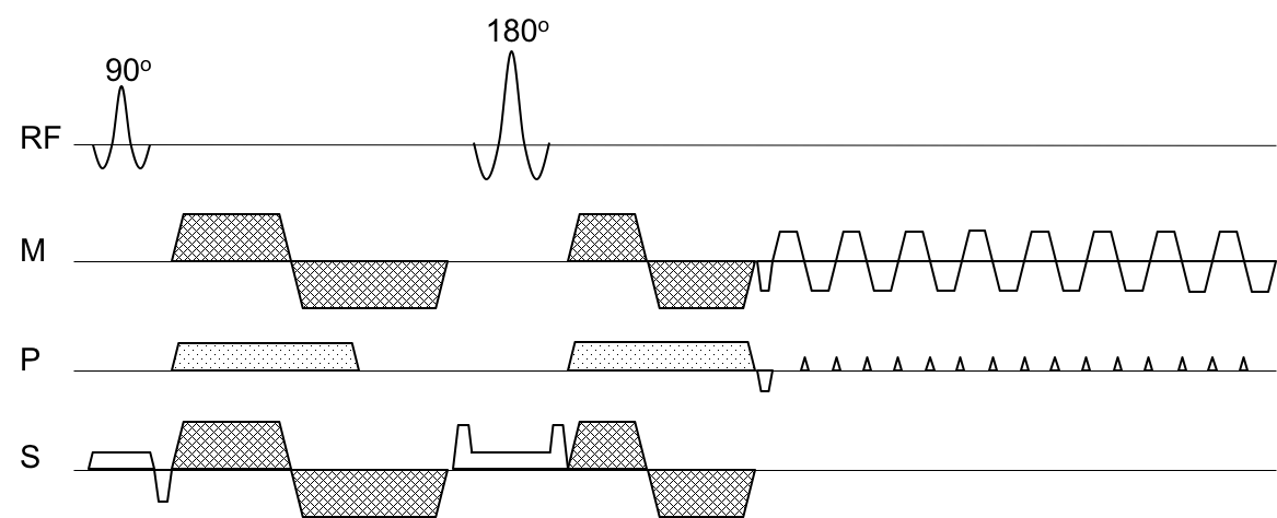
The proposed diffusion encoding combines an optimized asymmetric velocity-compensated diffusion encoding waveform (asym vc) on the M and S axes, and a traditional Stejskal-Tanner diffusion encoding (pgse) waveform on the P axis. The velocity-compensated diffusion waveform is asymmetric in order to reduce the echo time, and the duration of each gradient lobe was found by running a constrained optimization algorithm. Figure 1a shows the velocity compensated component of the proposed diffusion encoding waveform, which was assumed to have 2 lobes on either side of the $$$180°$$$pulse. It was also assumed that each lobe would ramp up to the maximum gradient strength at the maximum allowed slew rate. The constraints being applied to the optimization procedure, given in Figure 1c, were the M0 and M1 nulling, and the timing constraints for the 4 lobes. An expression for the b-value was found for this configuration of gradient lobes, and the echo time was minimized for a given target b-value. This gave the analytical expressions for each lobe plateau duration shown in Figure 1d. No correction was done for concomitant magnetic fields. Figure 2 shows the asym vc-pgsep waveform for all 3 axes.
MR measurement
In-vivo experiments were carried out in 5 subjects to assess the performance of the proposed asym vc-pgsep diffusion encoding scheme in motion resistance and vessel signal suppression. For comparison, experiments with pgse in all three axes and bipolar-vc in all three axes were also performed. Imaging parameters included: acquisition voxel size $$$=3x3x6mm^3$$$; 10 slices placed in the upper part of the liver; 3 orthogonal diffusion encoding directions at b-values of $$$[0,100,200,300,400]$$$s/mm2 and averages of $$$[4,4,5,6,8]$$$; TE=55/71/64ms for pgse, bipolar-vc and asym vc-pgsep; full Fourier encoding for pgse and a partial Fourier encoding factor of 0.7 for bipolar-vc and asym vc-pgsep. All experiments were performed on a 3T scanner (Philips Ingenia Elition, Best, The Netherlands) using anterior and posterior torso coils for receiving. ADC maps were calculated from b-values of $$$200,300$$$ and $$$400$$$ in order to avoid including the perfusion signal that can be detected at lower b-values.
Results
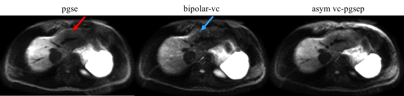
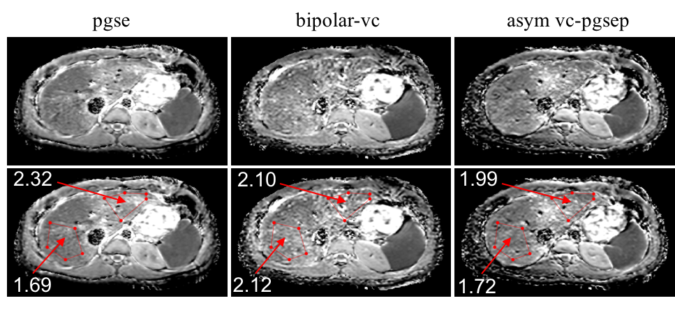
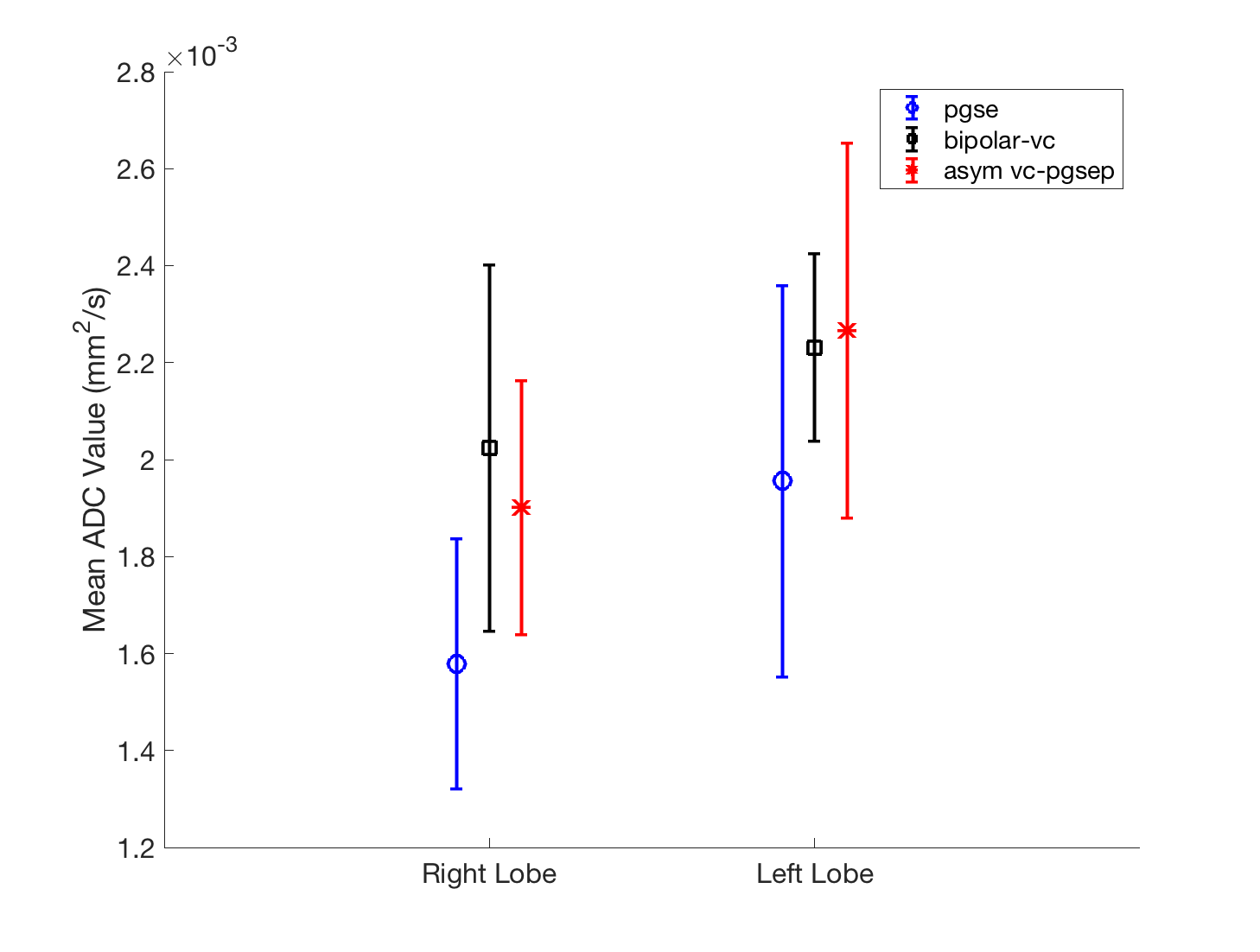
Figure 3 shows that bipolar-vc increases DW liver signal homogeneity in the left liver lobe but suffers from increased vessel signal throughout the liver. The proposed diffusion-encoding asym vc-pgsep maintains the left liver lobe signal in the high b-value images while providing good overall signal suppression. Figure 4 shows one case where asym vc-pgsep gave improved ADC quantification in the left liver lobe compared to pgse, and gave an ADC map with higher SNR and lower vessel signal compared to bipolar-vc. Figure 5 shows that across all subjects, bipolar-vc and asym vc-pgsep typically tended to give higher ADC values than pgse in the right liver lobe, which is not expected to be heavily affected by cardiac motion. The mean ADC values increased for all waveforms in the left liver lobe when compared to the right liver lobe.Discussion & Conclusion
The proposed partial optimized asymmetric velocity-compensated diffusion encoding, which combines traditional Stejskal-Tanner diffusion encoding in the phase encoding axis with optimized asymmetric velocity-compensated diffusion encoding in the frequency and slice encoding axes can improve vessel signal suppression and SNR compared to bipolar velocity-compensated diffusion encoding. However, the ADC values of any velocity-compensated acquisition are not directly comparable to the ADC values of a pgse acquisition. A larger scale study is needed to compare the performance of the different velocity compensated acquisition encoding waveforms to pgse acquisitions.Acknowledgements
The present work was supported by Philips Healthcare.References
1. Namimoto T, Yamashita Y, Sumi S, et al. Focal liver masses: characterization with diffusion-weighted echo-planar MR imaging. Radiology 1997; 204:739-744.
2. Ichikawa T, Haradome H, Hachiya J, et al. Diffusion-weighted MR imaging with a single-shot echoplanar sequence: detection and characterization of focal hepatic lesions. Am J Roentgenol 1998; 170:397-402.
3. Aliotta E, Wu HH, Ennis DB. Convex optimized diffusion encoding (CODE) gradient waveforms for minimum echo time and bulk motion compensated diffusion weighted MRI. Magn Reson Med 2017; 77:717–729
4. Peña-Nogales Ó, de Luis-Garcia R, Hernando D, et al. Optimal design of motion-compensated diffusion gradient waveforms. Proc. Intl. Soc. Mag. Reson. Med. 25 (2017) Abstract 3340
5. Peña-Nogales Ó, Zhang Y, Wang X, de Luis-Garcia R, Aja-Fernández S, Holmes J. H., Hernando D. Optimal Diffusion-weighting Gradient Waveform Design (ODGD): Formulation and Experimental Validation. Proc. Intl. Soc. Mag. Reson. Med. 26 (2018) Abstract 1615
6. Van AT, Cervantes B, Karampinos DC, et al. Partial velocity-compensated diffusion encoding for combined motion compensation and residual vessel signal suppression in liver DWI. Proc. Intl. Soc. Mag. Reson. Med. 26 (2018) Abstract 0079
7. Majeed W, Kalra P, Kolipaka A. Motion Compensated, Optimized Diffusion Encoding (MODE) Gradient Waveforms. Proc. Intl. Soc. Mag. Reson. Med. 26 (2018) Abstract 1614
8. Peña-Nogales Ó, Zhang Y, Wang X, de Luis-Garcia R, Aja-Fernández S, Holmes J. H., Hernando D. Optimized Diffusion-Weighting Gradient Waveform Design (ODGD) formulation for motion compensation and concomitant gradient nulling. Magn. Reson. Med. 2018;00:1–15. https://doi. org/10.1002/mrm.27462
9. Taouli B, Sandberg A, Stemmer A, et al. Diffusion-weighted imaging of the liver: comparison of navigator triggered and breathhold acquisitions. J Magn Reson Imaging 2009; 30:561-568.
10. Kwee TC, Takahara T, Niwa T, et al. Influence of cardiac motion on diffusion-weighted magnetic resonance imaging of the liver. Magn Reson Mater Phy 2009; 22:319-325.
11. Nasu K, Kuroki Y, Minami M. Feasibility of diffusion-weighted imaging under split breath-hold acquisition and postprocessing (DWI-SBAP): an attempt to suppress hepatic pseudo-anisotropy. Jpn J Radiol 2009; 27:78-850.
12. Ozaki M, Inoue Y, Miyati T, et al. Motion artifact reduction of diffusion-weighted MRI of the liver: use of velocity-compensated diffusion gradients combined with tetrahedral gradients. J Magn Reson Imaging 2013; 37:172-178.
13. Metens T, Absil J, Denolin V, et al. Liver apparent diffusion coefficient repeatability with individually predetermined optimal cardiac timing and artifact elimination by signal filtering. J Magn Reson Imaging 2016; 43:1100-1110.
14. Aliotta E, Wu HH, Ennis DB. Convex optimized diffusion encoding (CODE) gradient waveforms for minimum echo time and bulk motion-compensated diffusion-weighted MRI. Magn Reson Med 2017; 77:717-729.
15. Storey P, Frigo FJ, Hinks RS, Mock BJ, Collick BD, Baker N, Marmurek J, Graham SJ, Partial k-Space reconstruction in single-shot diffusion- weighted echo-planar imaging. Magn Reson Med 2007, 57:614-619.
16. Anderson AW, Gore JC, Analysis and correction of motion artifacts in diffusion-weighted imaging. Magn Reson Med 1994; 32:379-387. 17. Van AT, Karampinos DC, Georgiadis JG, Sutton BP, k-space and image-space combination for motion-induced phase-error correction in self-navigated multicoil multishot DWI. IEEE TMI 2009; 28:1770-1780.
Figures