1703
Volume-wise lesion detection on hepatic hemangioma and cyst considering inter-slice information through a 3D convolutional neural network1Philips Healthcare, Suzhou, China, 2Beijing Longleding Medical Technology Co., Ltd., Beijing, China
Synopsis
MR has been widely used for the diagnosis of hepatic hemangioma and cyst due to its significance of detection on small lesions. This study proposes a deep learning based method to detect the lesion volume in a three-dimensional manner on the dynamic contrast-enhanced MR images with hemangioma and/or cyst lesions. The results show good alignment of automated detection contour with the manually labelled lesion contour by professional radiologists, as well as accurate classification of lesion types.
Introduction
MR has been widely used for the diagnosis of hepatic hemangioma and cyst due to its significance of detection on small lesions [1]. However, the detection and delineation of the lesions were labor-intensive with manual labelling and it heavily relies on the clinical experience of the radiologist. The use of machine learning based method for lesion detection has been increasely reported, with a few studies using a 2D deep learning neural network [2], however, the 2D lesion ROIs may mislead some regions interpretation due to the lack of cross-slice information. In this study, we proposed a deep learning based method to detect the lesion volume in a three-dimensional manner on the dynamic contrast-enhanced MR images with hemangioma and/or cyst lesions.Method
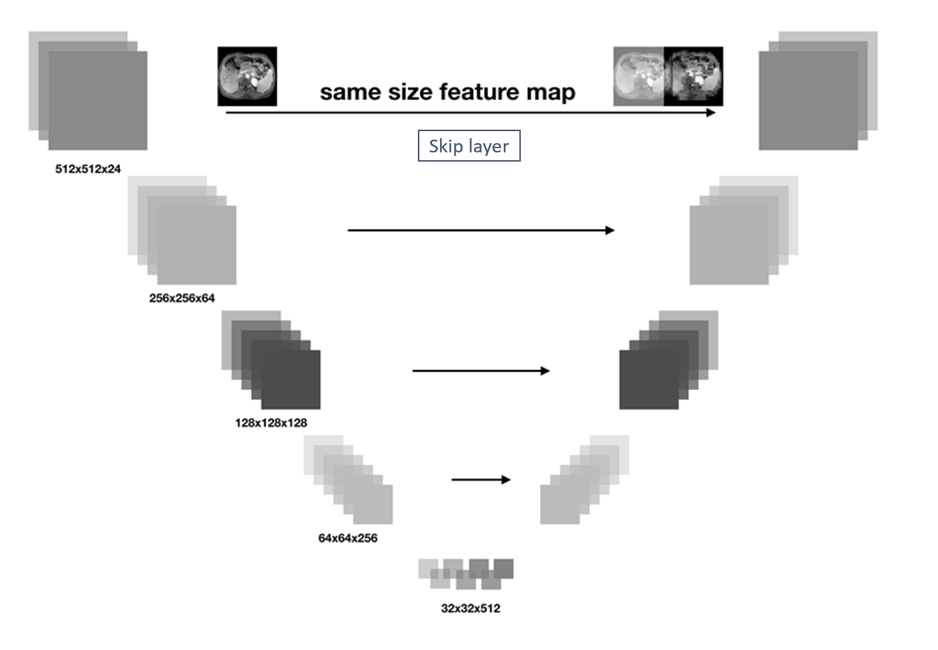
A cohort of 61 patients with either hepatic hemangioma or hepatic cyst were included in this study. Clinical routine dynamic contrast-enhanced (DCE) MR imaging were acquired on a 3T scanner (Philips Healthcare, the Netherlands). The delayed phase MR images were used to establish our deep learning neural network. Each patient data contains 40-45 slices depending on the coverage of the liver. Manual labeling of the lesions were performed by experienced radiologists with cross-check. To leverage the correlative information of lesion between consecutive slices, we employed a UNet neural network architecture with 3D Convolution kernels [3]. The UNet was a symmetric architecture in which the corresponding layer pairs in convolution and deconvolution steps had the same size of feature map and there was a skip layer structure between each pair as shown in Fig.1. The skip layer here passed the low level subtle details from original image (feature map) which promoted the segmentation result, while 3D kernels was preferred to 2D ones which could help to capture the inter-layer correlations of anatomical information that provided the lesion segmentation result a reasonable gain. The network was implemented through Tensorflow [4], with GPU 1.4GHz, 1080Ti. Original data were pre-processed by the DLTK software package. 80% of patient data were randomly arranged and used as training set and the rest as validation set.Results
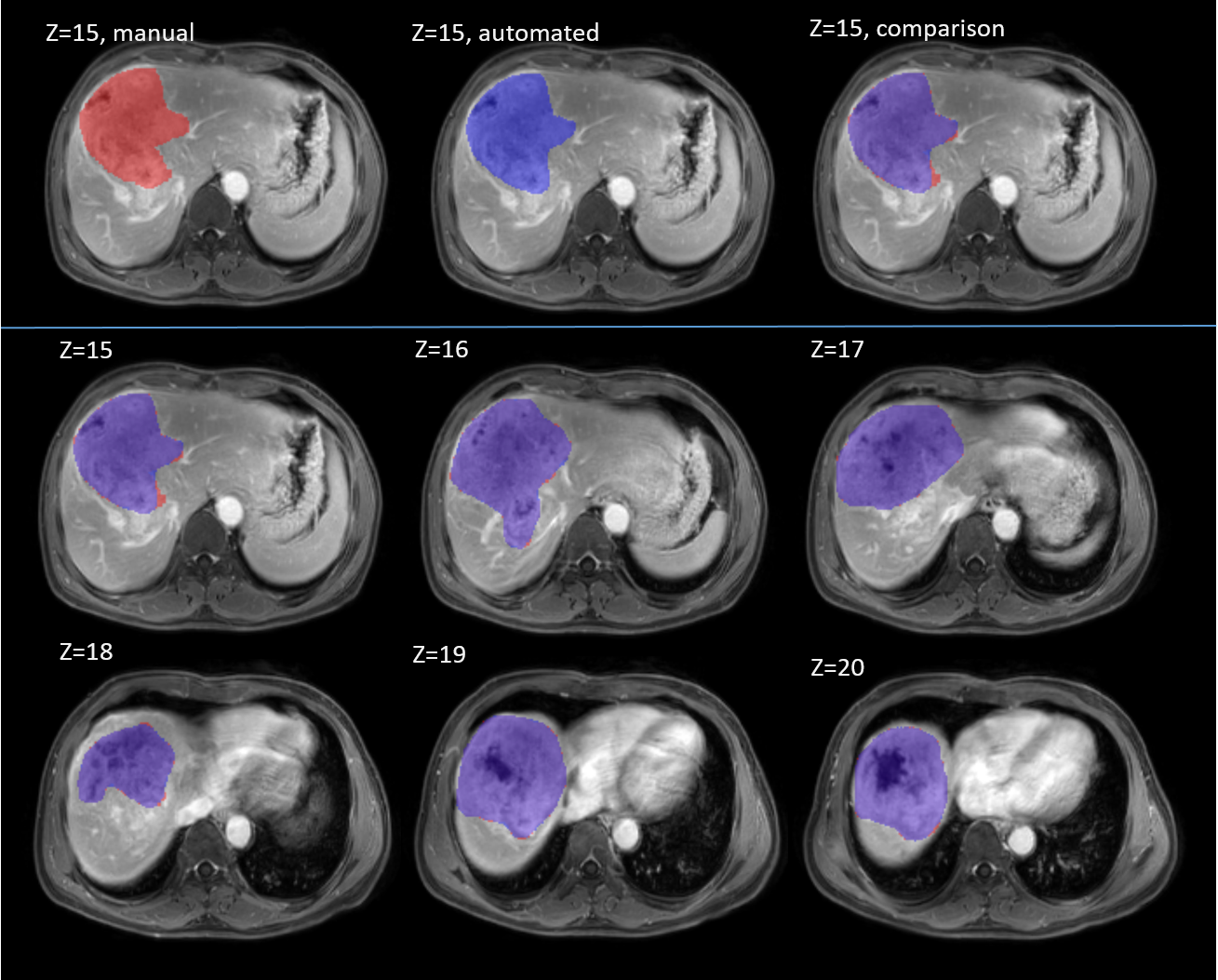
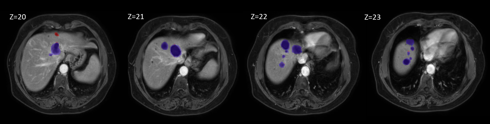
Figure 2 showed the comparison of lesion detection by manual delineation vs. 3D UNet segmentation. Region in red showed the manual labeled lesion, region in blue as the automated segmentation by our proposed method. Consecutive slices with the lesion contoured by manual delineation vs. by 3D UNet segmentation were shown. The region in purple indicated the overlap between the two labels. Dice overlap ratio for the whole 3D lesion volume was 91%. Similarly, Figure 3 displayed the comparison of 3D lesion volume detection versus the manual delineation, and the dice overlap ratio of 84% were obtained. For the validation set, the average dice overlap ratio could be 78%, indicating a good alignment of the detecting lesion contours.Discussion and Conclusion
In this study, we employed a 3D convolutional neural network with skip layer which takes inter-layer information and low-level details into consideration. The overlap ratio on a volume lesion between our result and the manual labelling were 84% for abovementioned dataset. Compared to the 2D convolutional network such as in [2], this network takes the inter-slice information into account, which potentially reduce the misleading of suspicious regions which may appear on isolated slice. As more emerging methods of semantic segmentation in the field of computer vision have been introduced to medical image segmentation, the current model could be further improved.
Acknowledgements
No acknowledgement found.References
[1] Sasaki K, Ito K, Koike S, et al. Differentiation between hepatic cyst and hemangioma: additive value of breath-hold, multisection fluid-attenuated inversion-recovery magnetic resonance imaging using half-Fourier acquisition single-shot turbo-spin-echo sequence. J Magn Reson Imaging. 2005;21: 29-36.
[2] Zhang Y, et al. An automated lesion detection method on hepatic hemangioma and hepatic cyst using fully convoluted network. ISMRM 2018.
[3] Erden B, Gamboa N, Wood S. 3D Convolutional Neural Network for Brain Tumor Segmentation. http://cs231n.stanford.edu/reports/2017/pdfs/526.pdf.
[4] Wongsuphasawat K, Smilkov D, Wexler J, et al. Visualizing Dataflow Graphs of Deep Learning Models in TensorFlow. IEEE Trans Vis Comput Graph. 2017;
Figures