1702
Noninvasive evaluation of the pathologic grade of hepatocellular carcinoma using MCF-3DCNN: A pilot studyDa-wei Yang1,2, Xiao-pei Wang1, Zheng-han Yang1, Zhen-chang Wang1, and Xi-bin Jia3
1Beijing friendship hospital, Capital medical university, Beijing, China, 2Beijing Key Laboratory of Translational Medicine on Liver Cirrhosis, Beijing, China, 3Beijing University of Technology, Beijing, China
Synopsis
This pilot study indicated that the MCF-3DCNN model may be valuable for the noninvasive evaluation of the pathologic grade of HCCs; however, further improvement would be necessary to achieve a better diagnostic performance for moderately and poorly differentiated HCCs.
Purpose
To evaluate the diagnostic performance of deep learning with a multichannel fusion three-dimensional convolutional neural network (MCF-3DCNN) in the differentiation of the pathologic grades of hepatocellular carcinoma (HCC) based on dynamic contrast-enhanced magnetic resonance images (DCE-MR images).Methods and Materials
Fifty-one histologically
proven HCCs from 42 consecutive patients from January 2015 to September 2017
were included in this retrospective study. Pathologic examinations revealed nine
well-differentiated, 35 moderately differentiated, and seven poorly
differentiated HCCs. DCE-MR images with five phases were collected using a 3.0
Tesla MR scanner. The 4D-tensor representation was employed to organize the collected
data in one temporal and three spatial dimensions by referring to the phases
and 3D scanning slices of the DCE-MR images. A deep learning diagnosis model
with MCF-3DCNN was proposed, and the structure of MCF-3DCNN was determined to approximate
clinical diagnosis experience by taking into account the significance of the spatial
and temporal information from DCE-MR images. Then, MCF-3DCNN was trained based
on well-labeled samples of HCC lesions from real patient cases by experienced
radiologists. The accuracy when differentiating the pathologic grades of HCC was
calculated, and the performance of MCF-3DCNN in lesion diagnosis was assessed.
Additionally, the areas under the receiver operating characteristic curves
(AUC) for distinguishing well-differentiated, moderately differentiated, and poorly
differentiated HCCs were calculated.Results
The average accuracy of the
gross differentiation of the pathologic grade of HCC via the MCF-3DCNN in the test data was 0.7396±0.0104, and the
average sensitivity and precision were 0.7396±0.0104 and 0.8042±0.0198, respectively. MCF-3DCNN also achieved the
highest diagnostic performance for discriminating well-differentiated HCCs from
others, with an average AUC, accuracy, sensitivity and specificity of 0.96,
91.00%, 96.88%, and 89.62%, respectively. Conclusions
This study indicates that MCF-3DCNN can be a promising technology for evaluating the pathologic grade of HCC based on DCE-MR images.Acknowledgements
The authors would like to express our enormous appreciation and gratitude to all participants.References
- Tang Y, Wang H, Ma L, et al. Diffusion-weighted imaging of hepatocellular carcinomas: a retrospective analysis of correlation between apparent diffusion coefficients and histological grade. Abdom Radiol 2016;41:1539-1545.
- Bruix J, Sherman M. Management of hepatocellular carcinoma. Hepatology 2005; 42:1208-1236.
- Kim DJ, Clark PJ, Heimbach J, et al. Recurrence of hepatocellular carcinoma: importance of mRECIST response to chemoembolization and tumor size. Am J Transplant 2014;14:1383-1390.
- Choi JW, Lee JM, Kim SJ, et al. Hepatocellular carcinoma: imaging patterns on gadoxetic acid-enhanced MR Images and their value as an imaging biomarker. Radiology 2013;267: 776-786.
- Castellano G, Bonilha L, Li LM, Cendes F. Texture analysis of medical images. Clin Radiol 2004;59:1061-1069.
Figures
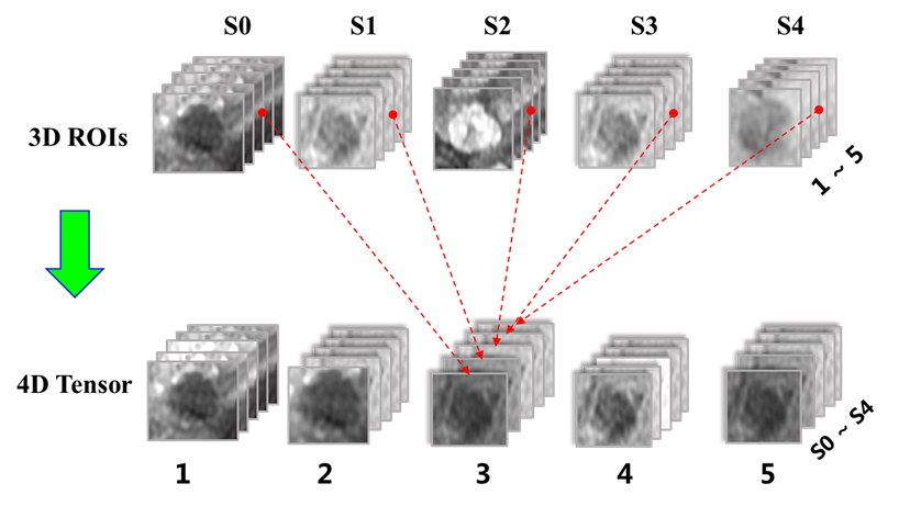
Figure 1 Data
representation with 4th-order tensor.
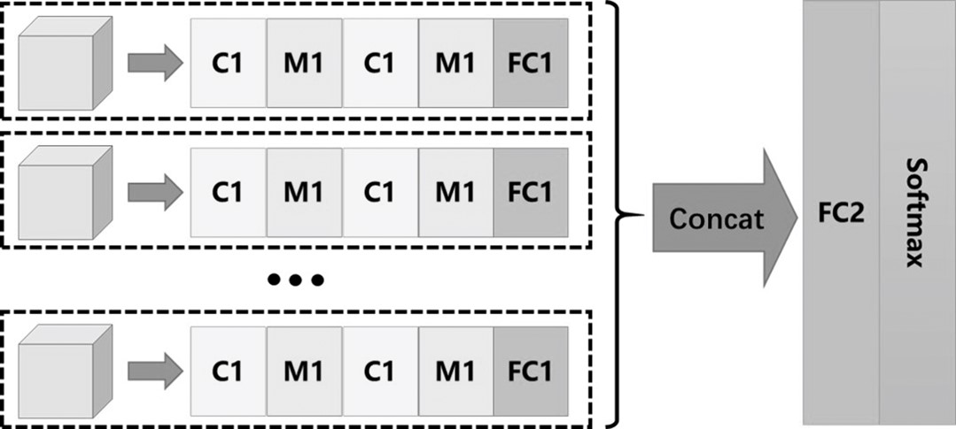
Figure 2 The architecture of the MCF-3D CNN.
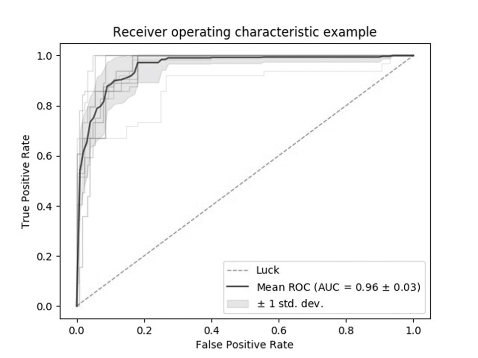
Figure 3
The average area under the ROC curve for 3DCNN for discriminating
well-differentiated HCCs from the others was 0.96.