1684
MR-based Radiomics Signature to Discriminate Different Pathologic Grade of Hepatocellular Carcinoma1The First Affiliated Hospital of Dalian Medical University, Dalian, China, 2Huiying Medical Technology Co., Beijing, China
Synopsis
Recently, the term radiomics (the extraction of multiple quantitative features from images) has drawn attention. Several cancer-related radiomics studies suggested that some quantitative imaging descriptors (such as texture features derived from MRI) could provide more information for cancer diagnosis. In the current study, MR-based radiomics signature was demonstrated to be capable to assess different pathologic grade of hepatocellular carcinoma, which will provide more prognostic information and facilitate clinical management.
Purpose
To investigate the application of MR-based radiomics signature to discriminate different pathologic grade of hepatocellular carcinoma.Introduction
Hepatocellular carcinoma (HCC) is the most common histologic type, accounting for more than 80%-90% of primary hepatic cancer, and it has caused high morbidity and mortality1. The degree of differentiation of HCC was demonstrated by numerous studies as a significant factor for prognosis. Poor differentiation of HCC would be a risk factor for tumor seeding or intrahepatic dissemination after treatment delivery2. Thus, accurate grading of HCC is vital for describe tumors’ biological behavior for prognosis and clinical decision. At present, the pathological analysis of tumor samples derived from surgery or puncture is a gold criteria to determine histological grade of HCC. Noninvasive and precise identification of HCC grade is extremely valuable for preoperative clinical practice. Radiomics, allowing a high throughput extraction of quantitative imaging features from tumors, has potential to perform better in assessing HCC differentiation based on MRI. Some studies have suggested that radiomics can be effective for grading tumors such as gliomas and bladder cancer3,4. Therefore, MR-based radiomics signature was introduced in the present study to evaluate its clinical application performance on HCC grade.Materials and Methods
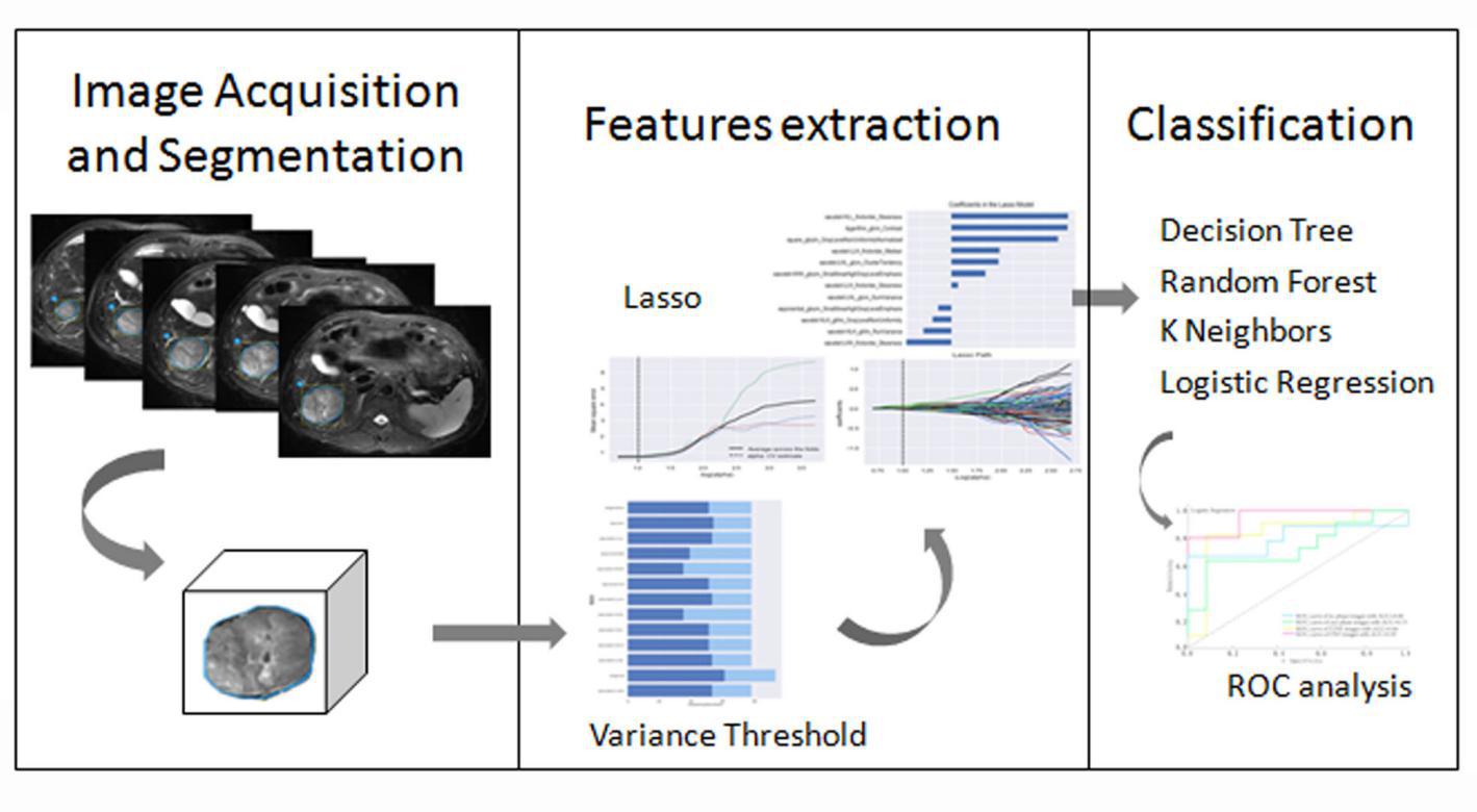
This retrospective study enrolled 166 patients who were pathologically confirmed as HCC, including 82 poorly differentiated HCCs and 84 non-poorly (well or moderately) differentiated HCCs. All patients have underwent preoperative MR examinations, including in-phase and out-phase T1WI, T2WI, DWI and LAVA dynamic contrast enhanced MRI (arterial, venous and delayed phase). The 80% samples were randomly selected as training set and the others were testing set. On the MR images, two radiologists manually outlined the ROIs which enclosed the boundary of target lesions and extracted 1029 radiomics features, which were classified as first order statistic, shape, gray level co-occurrence matrix (GLCM), gray level run length matrix (GLRLM) and gray level size zone matrix (GLSZM). Meanwhile, they were calculated by applying several filters such as exponential, square, square root, logarithm and wavelet with the exception of shape. Then, the variance threshold, select k best, and least absolute shrinkage and selection operator (LASSO) algorithms were explored for dimensionality reduction of the features. We used four classifiers ( logistic regression (LR), random forest (RF), decision tree (DT), k-nearest neighbors (KNN) ) to identify HCC grade on the basis of radiomics features. Diagnostic performance was evaluated by receiver operating characteristic (ROC) analysis.Results
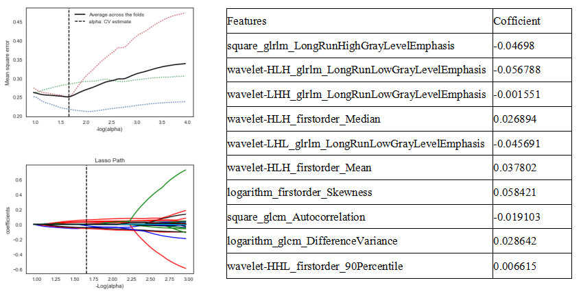
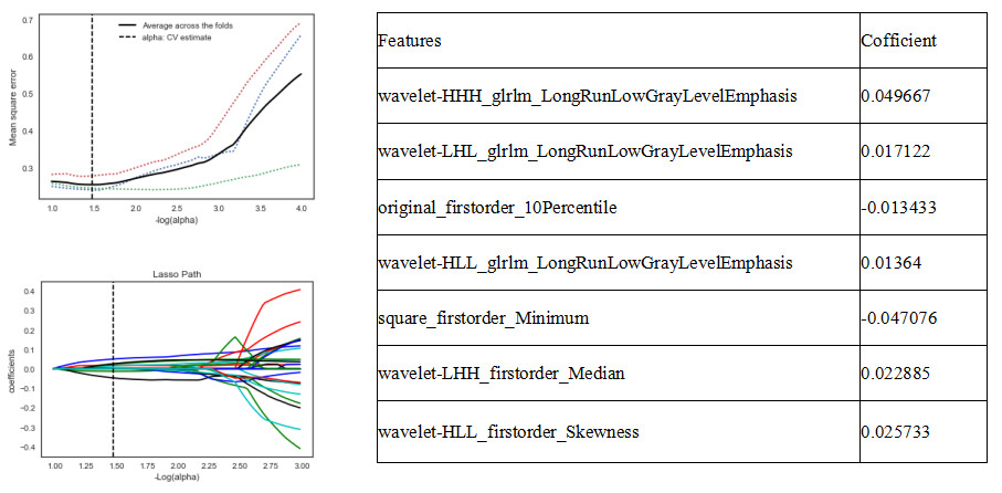
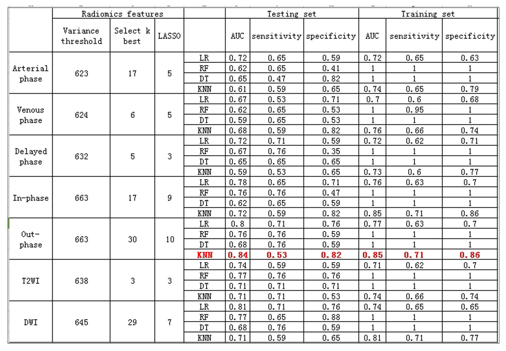
The valuable radiomics features which may quantitatively describe the HCC lesions, were selected on in-phase T1WI (n=9), out-phase T1WI (n=10), T2WI (n=3), DWI (n=7), arterial phase (n=5), venous phase (n=5) and delayed phase (n=3), respectively. Focusing on AUC which was the highest on each sequence, four classification strategy were present: KNN model on out-phase T1WI and venous images, LR model on in-phase T1WI, T2WI, DWI, arterial and delayed images. To identify poorly differentiated HCC, KNN classifier showed the best diagnostic performance based on radiomics features from out-phase T1WI images with a maximum AUC of 0.85 (sensitivity, 0.71; specificity, 0.86) in training set and AUC of 0.84 (sensitivity, 0.53; specificity, 0.82) in testing set. Results indicated that LR classifier on DWI images ( AUC: 0.74, sensitivity: 0.65, specificity: 0.65 in training set; AUC: 0.81, sensitivity: 0.71, specificity: 0.76 in testing set) was the optimal strategy to identify poorly differentiated HCC.Discussion
The radiomics based strategy has shown great potential in grading HCC, and out-phase T1WI and DWI images contain more useful cues of poorly differentiated HCC by using KNN and LR classifier, respectively. Recent studies5 have indicated that the signal intensity on DWI images was potential predictor of histological grade of HCC, which was also validated in our study. Discriminative features in the optimal feature subset were the GLRLM and first order of DWI, and the GLRLM, first order, and GLCM of out-phase T1WI. All these results suggest that GLRLM and first order features of DWI and out-phase T1WI images seem more important for accurate assessment of HCC grades.Conclusion
Poor differentiation of HCC has been proved as a risk factor of bad prognosis, and it can only be accurately identified by pathology. In this study, we proposed a MRI based radiomics strategy to preoperatively identify HCC grade, which will provide more prognostic information and facilitate clinical management.Acknowledgements
No acknowledgement found.References
[1] Omata M, Cheng AL, Kokudo N, et al. Asia-Pacific clinical practice guidelines on the management of hepatocellular carcinoma: a 2017 update. Hepatology International, 2017, 11(4): 317.
[2] Regimbeau J M, Abdalla E K, Vauthey J N, et al. Risk factors for early death due to recurrence after liver resection for hepatocellular carcinoma: Results of a multicenter study. Journal of Surgical Oncology, 2004, 85(1): 36-41.
[3] Kang Y, Choi S, Kim Y, et al. Gliomas: histogram analysis of apparent diffusion coefficient maps with standard- or high-b-value diffusionweighted MR imaging-correlation with tumor grade. Radiology, 2011, 261(3): 882–890.
[4] Zhang X, Xu X, Tian Q, et al. Radiomics assessment of bladder cancer grade using texture features from diffusion-weighted imaging. Journal of Magnetic Resonance Imaging Jmri, 2017,46(5): 1281-1288.
[5] Nasu K, Kuroki Y, Tsukamoto T, et al. Diffusion-weighted imaging of surgically resected hepatocellular carcinoma: Imaging characteristics and relationship among signal intensity, apparent diffusion coefficient, and histopathologic grade. American Journal of Roentgenology, 2009, 193(2): 438.
Figures