1679
Comparison between 3T MRI and CT for preoperative T staging of resectable esophageal cancer, with histopathological correlation1Radiology, Henan Cancer Hospital, Zhengzhou, China, 2MR Scientific Marketing, Siemens Healthcare, Shanghai, China
Synopsis
Seventy fourth patients with endoscopically proven EC and indeterminate T1/T2/T3/T4a stage by CT and EUS were enrolled prospectively. The diagnostic performances of MRI and CT were evaluated based on the sensibility, specificity and accuracy rate, the difference of accuracy rates between MRI and CT was analyzed by c2 test. This study showed MRI can obtain clear images of esophageal wall for preoperative T staging of EC with significantly higher accuracy rate than that of CT, and provide another high-accuracy non-invasive examination method for preoperative T staging of EC.
Introduction: Esophageal cancer (EC) is the eighth most common cancer (1). Staging of EC determines the selection of optimal treatment and predicts prognosis. Although endoscopic ultrasound (EUS) is considered as the most accurate method for the preoperative T staging of EC, but EUS is not recommended as a routine non-invasive method for preoperative examination of EC (2). In contrast, CT is a non-invasive technique, which is most commonly used in the preoperative T staging of EC. Because of the poor soft tissue resolution, conventional CT may not differentiate early tumor (T1 and T2 lesions), but is useful for the evaluation of tumor invasion into adjacent structures (3). Magnetic resonance imaging (MRI) with high soft tissue resolution is also non-invasive, however, the application of MRI in the chest examination has only recently be explored, largely due to technical improvements. The new technique BLADE with an acquisition scheme similar to the periodically rotated overlapping parallel lines with enhanced reconstruction can eliminate MR motion artifacts in brain and abdomen scans (4, 5), and Radial VIBE is also a new 3D gradient-echo sequence, which can acquire high quality images with improved lesion conspicuity in abdominopelvic MRI under free breathing (6). The purpose of our present study was to evaluate the image quality of T2-TSE-BLADE, DWI and radial VIBE in imaging of EC, and compare the diagnostic performance between MRI and CT in preoperative T staging of EC.
Purpose: To compare the T staging of resectable esophageal cancer (EC) using MRI and CT with that of the pathologic confirmation.
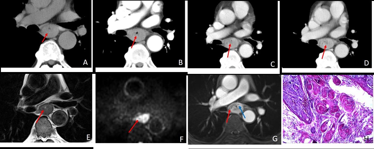
Materials and Methods: Seventy fourth patients with endoscopically proven EC and indeterminate T1/T2/T3/T4a stage by CT and EUS were enrolled prospectively. All patients underwent MRI and CT examinations 2 days after endoscopy. The sequences of MRI included T2-TSE-BLADE, diffusion-weighted imaging (DWI), and radial VIBE, CT were performed by plain and dynamic contrast enhanced scanning, and surgery was performed within one week after imaging. Two independent readers evaluated the MR image quality using a 5-point score and assigned the T staging. Two other CT readers evaluated the CT T staging, and post-operative pathologic confirmation was considered the gold standard. The diagnostic performances of MRI and CT were evaluated based on the sensibility, specificity and accuracy rate, the difference of accuracy rates between MRI and CT was analyzed by χ2 test.
Results:74 individuals were finally enrolled in the study (mean age, 60.4±8.2 years; range, 43-79 years; 56 men, 18 women), including 63 cases of squamous cell carcinoma, 7 cases of adenocarcinoma and 4 cases of neuroendocrine carcinoma confirmed by post-operative pathology. 22 cases were stage T1, 23 cases were stage T2, 24 cases were stage T3, and 5 cases were stage T4a. MRI Cases with better than average image quality were 60/74 by both readers, with good inter-reader agreement (Kappa=0.727, P<0.001). The consistency of MRI T staging was 82.4% (61/74, Kappa=0.807, P<0.001) with accuracy ranging between 90.5% and 95.9%, and the T staging consistency between CT and pathology was 60.8% (45/74, Kappa=0.441, P<0.001), with accuracy ranged between 74.3% and 93.2%. The difference of T staging accuracy rates between MRI and CT was statistically significant (χ2=12.5, P<0.05).
Conclusion: MRI has better than average image quality and showed advantage over CT for preoperative T staging of EC. Keywords: Esophageal cancer, Tumor staging, Magnetic resonance imaging, Computer tomography
Discussion: This study demonstrated that MRI can obtain high quality images of esophageal wall, and provide higher accuracy of preoperative T staging in EC than CT. MRI has several advantages in multi-sequence imaging and soft tissue resolution. When applied to a tubular structure such as the rectum (7), MRI can acquire high accuracy in evaluating depth of lesion infiltration. However, motion artifacts have led to the limitation of MRI in chest examination. Our study was the first time to accuracy of the combined T2-TSE-BLADE, DWI and radial VIBE together and then compare this accuracy with that of CT in T staging of EC. The results showed that MRI has higher accuracy than CT in T staging of EC. As reported by both readers, image quality of MRI was good enough for staging, and the inter-reader agreement was good. Conclusion: MRI can obtain clear images of esophageal wall for preoperative T staging of EC with significantly higher accuracy rate than that of CT, and provide another high-accuracy non-invasive examination method for preoperative T staging of EC.
Acknowledgements
No acknowledgement found.References
1. Heethuis SE, van Rossum PS, Lips IM, et al. Dynamic contrast-enhanced MRI for treatment response assessment in patients with oesophageal cancer receiving neoadjuvant chemoradiotherapy. Radiotherapy and oncology : journal of the European Society for Therapeutic Radiology and Oncology. 2016;120(1):128-35.
2. Schreurs LM, Janssens AC, Groen H, et al. Value of EUS in Determining Curative Resectability in Reference to CT and FDG-PET: The Optimal Sequence in Preoperative Staging of Esophageal Cancer? Annals of Surgical Oncology. 2011;23(5):1021-8.
3. Hong SJ, Kim TJ, Nam KB, et al. New TNM staging system for esophageal cancer: what chest radiologists need to know. Radiographics A Review Publication of the Radiological Society of North America Inc. 2014;34(6):1722-40.
4. Deng J, Miller FH, Salem R, Omary RA, Larson AC. Multishot diffusion-weighted PROPELLER magnetic resonance imaging of the abdomen. Investigative Radiology. 2006;41(10):769-75.
5. Lavdas E, Mavroidis P, Kostopoulos S, et al. Improvement of image quality using BLADE sequences in brain MR imaging. Magn Reson Imaging. 2013;31(2):189-200.
6. Chandarana H, Feng L, Block TK, et al. Free-breathing contrast-enhanced multiphase MRI of the liver using a combination of compressed sensing, parallel imaging, and golden-angle radial sampling. Investigative Radiology. 2013;48(1):10-6.
7. Kong M, Hong SE, Choi WS, Kim SY, Choi J. Preoperative Concurrent Chemoradiotherapy for Locally Advanced Rectal Cancer: Treatment Outcomes and Analysis of Prognostic Factors. Cancer Research & Treatment Official Journal of Korean Cancer Association. 2012;44(2):104-12.
Figures