1668
Usefulness of amide proton transfer imaging in the evaluation of autoimmune pancreatitis activity1Kagoshima University Graduate School of Medical and Dental Sciences, kagoshima, Japan, 2Department of Radiological Technology, Kagoshima University Hospital, Kagoshima, Japan, 3Philips GmbH Innovative Technologies Research Laboratories, Hamburg, Germany, 4Philips Electronics Japan, Tokyo, Japan
Synopsis
This study focused on the potential of amide proton transfer (APT) MR imaging at 3.0T as an objective imaging biomarker in patients with autoimmune pancreatitis (AIP). Correlation of serum immunoglobulin G4 (IgG4) levels with APT SI, the apparent diffusion coefficient (ADC) values or the maximum standardized uptake value (SUVmax) on 18F-fluorodeoxyglucose positron emission tomography (18F-FDG PET) was evaluated in eleven patients with AIP. Our results showed a significant positive correlation between serum IgG4 levels and APT signal intensity (SI). Therefore, APT imaging might be useful for monitoring AIP activity.
Introduction
Autoimmune pancreatitis (AIP) is a type of pancreatitis for which an immune-mediated mechanism has been postulated. AIP is characterized by mixed inflammatory cell infiltrates centered around the pancreatic ducts in association with tumor-like regions of fibrosis, leading to organ dysfunction. It is important not only to diagnose AIP but also to rule out active or recurrent inflammation in the long-term follow-up of AIP patients. Symptoms at early active or early recurrent stage are not necessarily specific. Serum immunoglobulin G4 (IgG4) is closely associated with AIP activity,1,2 but the affected organs cannot be detected with serological findings alone. Therefore, objective imaging biomarker might be useful for early therapeutic intervention.
Diffusion-weighted (DW) imaging is based on tissue microstructure induced changes in the Brownian motion of water molecules. Through the measurement of apparent diffusion coefficient (ADC), DW imaging provides qualitative and quantitative information that is pertinent to tissue structure. Amide proton transfer (APT) MR imaging is a type of chemical exchange saturation transfer imaging which specifically probes intrinsic amides, i.e., proteins and peptides.3 No attempts have been made to apply APT imaging to AIP activity.
Purpose
The purpose of this study was to determine whether APT imaging can be used as an objective biomarker for AIP activity.Methods
Our study population consisted of 11 consecutive patients (9 men and 2 women; mean age, 67.0 years; age range, 54–79 years) with autoimmune pancreatitis, who underwent DW (b=0 and 1000 sec/mm2) and APT imaging at 3.0T before treatment. APT imaging data were acquired in a coronal plane using a respiratory-triggered single-slice turbo-spin-echo sequence with a saturation power level of 2 μT and a duration of 1.7 s at 7 saturation frequency offsets -5 to +5 ppm with a step of 1.5 ppm as well as one far-off-resonant frequency (-1560 ppm) for signal normalization. Imaging parameters were as follows: field of view, 500 × 500 mm2; voxel size, 2.2 × 2.2 mm2; slice thickness, 5 mm; repetition time, 1852 (respiratory intervals) ms; echo time, 4.4 ms; echo train length, 32; number of signal averages, 1; sensitivity encoding factor, 1. δB0 maps were separately acquired for a point-by-point δB0 correction. APT signal intensity (SI) was defined as magnetization transfer ratio asymmetry (MTRasym) at 3.5 ppm: MTRasym = (S [-3.5 ppm] – S [+3.5 ppm]) / S0. The total scan time of APT imaging was about 3 minutes.
Spearman’s bivariate correlation was used to assess the correlation of serum immunoglobulin G4 (IgG4) levels with APT SI, the apparent diffusion coefficient (ADC) values or the maximum standardized uptake value (SUVmax) on 18F-fluorodeoxyglucose positron emission tomography (18F-FDG PET).
Results
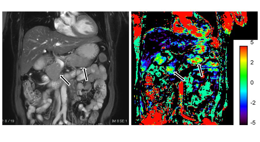
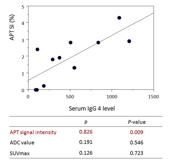
The mean APT SI of AIP was 1.9 ± 1.3% ranging from 0 to 4.3% (Fig. 1). There was a significant positive correlation between serum IgG4 and APT SI (P = 0.009, ρ= 0.826) (Fig. 2). No significant correlation was obtained between the serum IgG4 and ADC values (P = 0.546, ρ= 0.191) or SUVmax (P = 0.723, ρ = 0.126).Discussion
Our study demonstrated a significant positive correlation between the serum IgG4 levels and APT SI. Previous researchers reported the usefulness of DW imaging or 18F-FDG PET for monitoring AIP activity and the effects of steroid treatment.4,5 In our study, no significant correlation was obtained between the serum IgG4 and ADC values or SUVmax. Therefore, APT imaging might be more useful for evaluating AIP activity than DW imaging or 18F-FDG PET.Conclusion
APT imaging might be useful for evaluating AIP activity and the effect of steroid treatment.
Acknowledgements
No acknowledgement found.References
- Kamisawa T, Okazaki K, Kawa S, et al. Amendment of the Japanese consensus guidelines for autoimmune pancreatitis, 2013 III. Treatment and prognosis of autoimmune pancreatitis. J Gastroenterol. 2014;49:961-970.
- Kamisawa T, Shimosegawa T, Okazaki K, et al. Standard steroid treatment for autoimmune pancreatitis. Gut. 2009;58:1504-1507.
- Zhou J, Payen JF, Wilson DA, et al. Using the amide proton signals of intracellular proteins and peptides to detect pH effects in MRI. Nat Med. 2003;9:1085-1090.
- Taniguchi T, Kobayashi H, Nishikawa K, et al. Diffusion-weighted magnetic resonance imaging in autoimmune pancreatitis. Jpn J Radiol. 2009;27:138-142.
- Nakajo M, Jinnouchi S, Fukukura Y, et al. The efficacy of whole-body FDG-PET or PET/CT for autoimmune pancreatitis and associated extrapancreatic autoimmune lesions. Eur J Nucl Med Mol Imaging. 2007;34:2088-2095.
Figures