1662
Computer-aided pancreas segmentation based on 3D GRE Dixon MRIChao Ma1, Xiaoliang Gong2, Panpan Yang1, Yufei Chen2, Chaolin Du2, Caixia Fu3, Xu Yan4, and Jianping Lu1
1Radiology, Changhai Hospital of Shanghai, Shanghai, China, 2Country Key Laboratory of Embedded System and Service Computing (Ministry of Education), Tongji University, Shanghai, China, 3Siemens Shenzhen Magnetic Resonance Ltd., Shenzhen, China, 4Siemens Healthcare, Shanghai, China
Synopsis
Pancreas segmentation is of great significance for pancreatic cancer radiotherapy positioning, pancreatic structure and function evaluation, etc. In the study, we purposed a simple computer-aided pancreas segmentation method based on 3D GRE Dixon images by using a free open source software system.
INTRODUCTION
Computer aided detection/diagnosis (CAD) has important clinical application prospects. Specifically, the pancreas automatic segmentation is of great significance for pancreatic cancer radiotherapy positioning 1, structure and function of pancreatic evaluation of pancreas 2. In the study, we prospectively investigated the feasibility of computer-aided pancreas segmentation based on a threshold method and a morphological method by using 3D GRE Dixon images.METHODS
Seventeen volunteers underwent 3D routine and optimized Dixon MRI at 3.0 T whole body system (MAGNETOM Skyra, Siemens, Erlangen, Germany) in the current study. Four phase images including opposed phase, in-phase images, water images and fat images were reconstructed inline. The optimized 3D Dixon scan parameters are: recovery time TR, 3.92 ms; the echo time TE = 1.23/2.46 ms; the acquisition matrix 288 x 188; FOV 400 × 325 mm2; slice number 40; slice thickness 3 mm; slice gap 0 mm; the angle of 9 °. The automatic segmentation of pancreas is based on the Medical Imaging Interaction ToolKit (MITK) with radiologists selected images of oppose phase and water phase of two Dixon scans. Dice coefficients were calculated and compared for computer-aided pancreas segmentation based on the images of routine and optimized Dixon sequences.RESULTS
The dice coefficients of the computer-aided pancreas segmentation were 0.633±0.080 and 0.716±0.033 for opposed-phase and water images of routine Dixon MRI, while they were 0.415±0.143 and 0.779±0.048 for the optimized Dixon MRI. The Dice index was significantly higher based on the water images of optimized Dixon than that on the other three groups (all p values < 0.001).DISCUSSION
Previous study reported computer-assisted pancreas segmentation based on 2D or 3D CT images, but the accuracy of pancreas segmentation is significantly lower than that of other abdominal organs 3, the main reason is that the pancreas located in the depths of upper abdomen, and its surrounding tissues are relatively complex. In addition, the pancreas position varied with different individuals. MRI is an important tool to detect and characterize pancreatic diseases, with good soft tissue contrast superior to CT. Studies on the computer aided pancreas segmentation by using MRI images have been reported, such as Gou s. et al. explored computer-aided pancreas segmentation based on the 2D dynamic enhanced MRI 4 and 3D fat suppression VIBE MRI 5. Their newest results shown the acquired Dice index higher than 0.83 from 3 patients and 2 healthy volunteers’ total 12 imaging volumes. The method of automatic segmentation of abdominal organs based on the fat images from 3D Dixon sequence was proposed by Jun Shen et al 2, and the average Dice coefficient for 20 obese patients was 0.672. In the current study, based on the water image of the optimized Dixon, the automatic pancreas segmentation using our method obtained excellent similarity (the mean Dice coefficient was 0.779). It is noticed that the segmentation algorithm used in this study was based on the traditional threshold and morphological method. The optimized Dixon water images could obtain more stable segmentation results.CONCLUSION
Computer-aided pancreas segmentation based on the optimized Dixon MRI is feasible. The water images of Dixon obtained the best similarity.Acknowledgements
This work was supported by the project of science and technology innovation action (174119552200); Natural Science Foundation of Shanghai (16JC1401300); Natural Science Foundation of China (81601468, 61573235), project of precision medical transformation application of SMMU (2017JZ42).References
- Crijns SP, Raaymakers BW, Lagendijk JJ. Proof of concept of MRI guided tracked radiation delivery: Tracking one-dimensional motion. Phys Med Biol, 2012,57(23):7863-7872.
- Shen J, Baum T, Cordes C, et al. Automatic segmentation of abdominal organs and adipose tissue compartments in water-fat MRI: Application to weight-loss in obesity. Eur J Radiol, 2016, 85(9):1613-1621.
- Chu C, Oda M1, Kitasaka T, et al. Multi-organ segmentation based on spatially-divided probabilistic atlas from 3D abdominal CT images. Med Image Comput Comput Assist Interv 2013; 16:165-172. 4
- Gou S, Wu J, Liu F, et al. Feasibility of automated pancreas segmentation based on dynamic MRI. Br J Radiol 2014; 87:20140248.
- Gou S, Lee P, Hu P, et al. Feasibility of automated 3-dimensional magnetic resonance imaging pancreas segmentation. Adv Radiat Oncol 2016; 1:182-193.
Figures
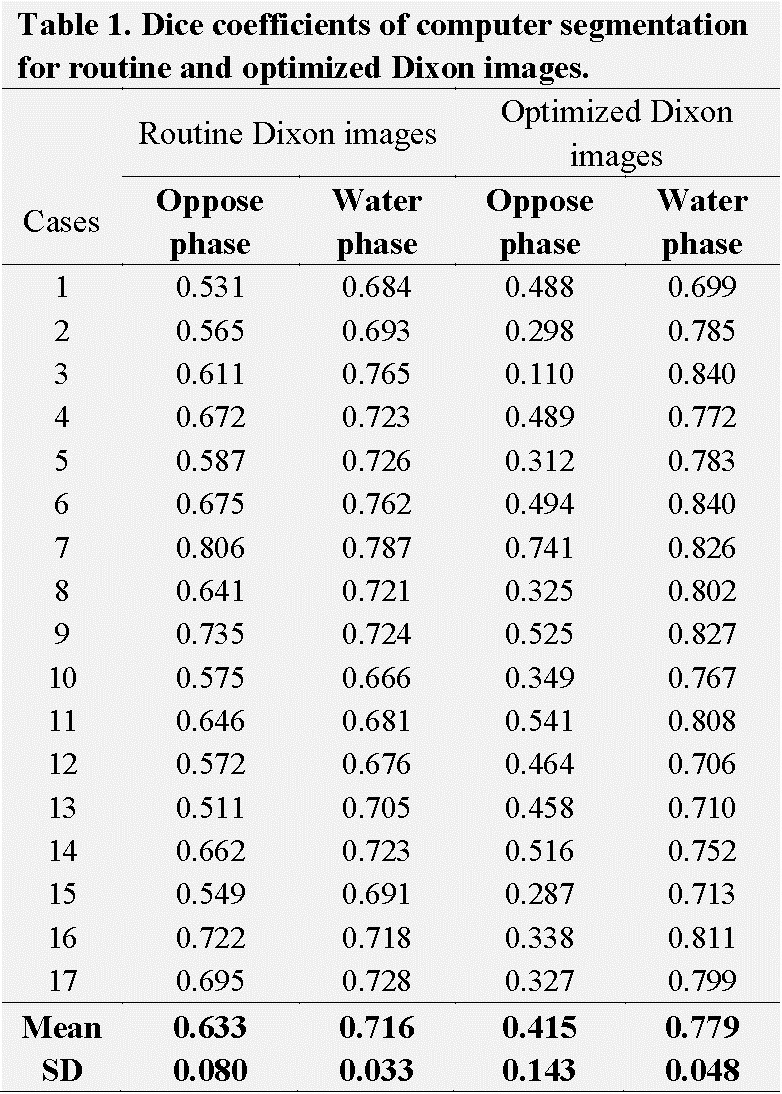
Table 1. Dice coefficients of computer segmentation for routine and optimized Dixon
images.
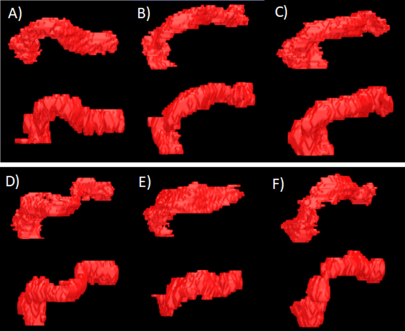
Figure 1.
3D Segmentation results by automatic segmentation
and manual ground truth of different ages respectively based on optimized Dixon
water phase images. (A) DI: 0.811; (B) DI: 0.808; (C) DI: 0.826; (D) DI: 0.802;
(E) DI: 0.772; (F) DI: 0.799.