1657
Slip-interface imaging preoperatively predicts hepatocellular carcinoma microvascular invasion1Department of Radiology, the Third Affiliated Hospital, Sun Yat-sen University (SYSU), Guangzhou, China, 2Department of Radiology, Mayo Clinic, Rochester, MN, United States
Synopsis
Hepatocellular carcinoma (HCC) is the most common type of primary liver cancer in adults. One of the most strongly correlated factors predicting outcome is the presence or absence of vascular invasion. Since microvascular invasion cannot be found with conventional CT or MRI examination, we investigated whether slip-interface imaging (SII) could identify HCC microvascular invasion. The results showed that in 32 of 33 patients with HCC, SII-assessed microvascular invasion agreed with pathology, indicating that this technique may become useful for detecting HCC microvascular invasion and guiding treatment planning.
Introduction
Hepatocellular carcinoma (HCC) is the most common type of primary liver cancer in adults and is the most common cause of death in people with cirrhosis [1,2]. Several factors predicting outcome have been identified including tumor characteristics such as size, stage, grade, the presence of vascular invasion, portal vein tumor thrombus, intrahepatic metastases, hepatitis status, functional liver reserve and the serum α-fetoprotein level. Overall, one of the most strongly correlated factors is the presence or absence of vascular invasion [3,4]. MR elastography (MRE) is a noninvasive, quantitative method that can detect changes in tissue biomechanics, such as tissue stiffness, brought about by disease [5,6]. Since many diseases cause substantial changes in the mechanical properties of tissue, this provides motivation for continuing to develop this technology to assess as many of these tissue properties as possible. The aim of this study was to investigate the ability of slip-interface imaging (SII), a recently developed MRE-based technique, to predict HCC microvascular invasion, using findings at pathology as the reference standard.Materials and Methods
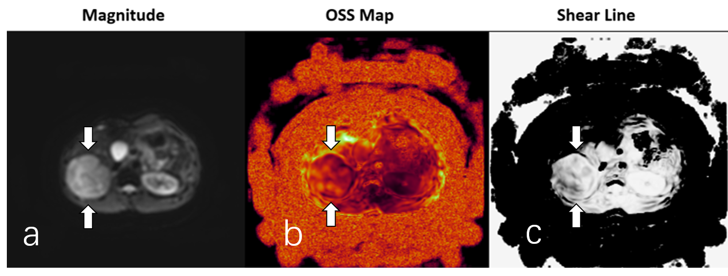
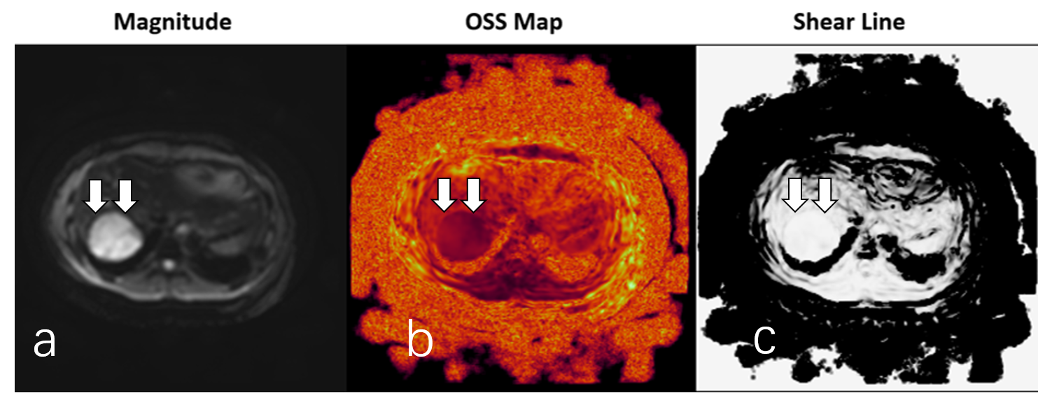
With Institutional Review Board approval and written informed consent, 33 patients from Oct. 2014 to May 2017 with HCC underwent preoperative SII assessment. MRE was performed on a 3.0T scanner (GE, Discovery MR750, Waukesha, WI) with a breath-held, multislice, single-shot, flow-compensated, spin-echo, EPI, 3D-MRE sequence using 60-Hz vibrations with the following imaging parameters: TR = 1334 ms, TE = 51.7-52.8 ms, slice thickness = 3.6 mm, 32 slices, bandwidth = 500 kHz, matrix= 256 × 96, 3 MRE phase offsets, 6 motion encoding directions, and acceleration factor of 2. An abdominal wall passive driver, developed by Mayo Clinic, was positioned over the upper abdominal wall and secured with an elastic belt. The MRE sequence was performed before the use of any intravenous contrast. On the scanner, an inversion algorithm automatically processed the raw data and produced grayscale and colorized stiffness maps (elastograms). The displacement data were reprocessed off-line to generate additional images reflecting the octahedral shear strain (OSS) and the presence of shear lines that were analyzed to determine microvascular invasion by inspecting these values along the interface of the tumor. The SII findings from the shear line images and OSS were independently and blindly correlated with pathology findings of microvascular invasion by using the Cohen's κ coefficient and chi‐squared test. Statistical significance was defined as P<0.05.Results
A pathologist categorized the pathological specimens as being without microvascular invasion in 13 patients, and with microvascular invasion in 20 patients. Both shear line images and OSS agreed with the pathological findings in 32 (96.97%) cases (Figures 1 and 2). The correlations between the SII and OSS predictions and the pathology findings were statistically significant (chi‐squared test, P = 0.01 and P < 0.001, respectively).Discussion
MRE is a noninvasive, quantitative method that can detect changes in tissue biomechanics, such as stiffness, brought about by disease, such as cancer and fibrosis [7-16]. Conventional imaging techniques, such as CT and MRI, can detect tumor size and macrovascular invasion, but cannot detect microvascular invasion. Our results show that SII and OSS derived from MRE images agreed well with the pathological findings. Our study demonstrated that MRE has the potential to provide HCC microvascular invasion information that may be useful for predicting therapeutic response preoperatively and noninvasively.Conclusion
SII is capable of preoperatively evaluating the presence of HCC microvascular invasion noninvasively, allowing for improved prediction of surgical risk and tumor resectability.Acknowledgements
81271562 (to J.W.)/National Natural Science Foundation of China 81271562/National Natural Science Foundation of China 01704020016 (to J.W.)/Science and Technology Program of Guangzhou, China EB001981 (to R.L.E.)/National Institutes of Health (NIH)EB017197 (to M.Y.)/National Institutes of Health (NIH) 201704020016/Science and Technology Program of GuangzhouReferences
[1] Forner A, Llovet JM, Bruix J. Hepatocellular carcinoma. Lancet, 2012, 379(9822): 1245-1255.
[2] Forner A, Reig M, Bruix J. Hepatocellular carcinoma. Lancet, 2018, 391(10127): 1301-1314.
[3] Griffin N, Addley H, Sala E, et al. Vascular invasion in hepatocellular carcinoma: is there a correlation with MRI? Br J Radiol, 2012, 85(1014): 736-744.
[4] Hsieh CH, Wei CK, Yin WY, et al. Vascular invasion affects survival in early hepatocellular carcinoma. Mol Clin Oncol, 2015, 3(1): 252-256.
[5] Pepin KM, Ehman RL, McGee KP. Magnetic resonance elastography (MRE) in cancer: Technique, analysis, and applications. Prog Nucl Magn Reson Spectrosc, 2015, 90-91: 32-48.
[6] Venkatesh SK, Ehman RL. Magnetic resonance elastography of abdomen. Abdom Imaging, 2015, 40(4): 745-759.
[7] Ichikawa S, Motosugi U, Ichikawa T, et al. Magnetic resonance elastography for staging liver fibrosis in chronic hepatitis C. Magn Reson Med Sci, 2012, 11(4): 291-297.
[8] Singh S, Venkatesh SK, Loomba R, et al. Magnetic resonance elastography for staging liver fibrosis in non-alcoholic fatty liver disease: a diagnostic accuracy systematic review and individual participant data pooled analysis. Eur Radiol, 2016, 26(5): 1431-1440.
[9] Venkatesh SK, Wang G, Lim SG, et al. Magnetic resonance elastography for the detection and staging of liver fibrosis in chronic hepatitis B. Eur Radiol, 2014, 24(1): 70-78.
[10] Herzka DA, Kotys MS, Sinkus R, et al. Magnetic resonance elastography in the liver at 3 Tesla using a second harmonic approach. Magn Reson Med, 2009, 62(2): 284-291.
[11] Morisaka H, Motosugi U, Ichikawa S, et al. Magnetic resonance elastography is as accurate as liver biopsy for liver fibrosis staging. J Magn Reson Imaging, 2018, 47(5): 1268-1275.
[12] Cui J, Heba E, Hernandez C, et al. Magnetic resonance elastography is superior to acoustic radiation force impulse for the Diagnosis of fibrosis in patients with biopsy-proven nonalcoholic fatty liver disease: A prospective study. Hepatology, 2016, 63(2): 453-461.
[13] Schwimmer JB, Behling C, Angeles JE, et al. Magnetic resonance elastography measured shear stiffness as a biomarker of fibrosis in pediatric nonalcoholic fatty liver disease. Hepatology, 2017, 66(5): 1474-1485.
[14] Loomba R, Wolfson T, Ang B, et al. Magnetic resonance elastography predicts advanced fibrosis in patients with nonalcoholic fatty liver disease: a prospective study. Hepatology, 2014, 60(6): 1920-1928.
[15] Wang J, Shan Q, Liu Y, et al. 3D MR Elastography of Hepatocellular Carcinomas as a Potential Biomarker for Predicting Tumor Recurrence. J Magn Reson Imaging, 2018.
[16] Wang J, Glaser KJ, Zhang T, et al. Assessment of advanced hepatic MR elastography methods for susceptibility artifact suppression in clinical patients. J Magn Reson Imaging, 2018, 47(4): 976-987.
Figures