1644
Amide proton transfer imaging for rectal cancer: correlation with IVIM, DCE MRI and 18F-FDG-PET/CT1Kagoshima University, Kagoshima, Japan, 2Philips GmbH Innovative Technologies, Research Laboratories, Hamburg, Germany, 3Philips Electronics, Japan, Tokyo, Japan
Synopsis
This study focused on the correlation between amide proton transfer
(APT) imaging and IVIM, DCE MRI or 18F-FDG PET/CT in rectal cancer. Our
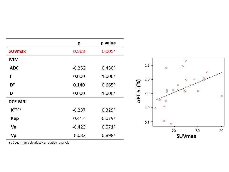
results showed a significant positive
correlation between APT signal intensity (APT SI) and SUVmax (p = 0.005, ρ = 0.547). No
significant correlation was shown between APT SI and IVIM (ADC, f, D* or D) or
DCE MRI parameters (Ktrans, Kep, Ve or
Vp).
These results suggested that APT imaging reflects some metabolism of the rectal
cancer and may be useful for response prediction after chemotherapy.
Introduction
The current trend in the treatment of rectal cancer is a more widespread acceptance of neoadjuvant therapies. However, there is concern that the opportunity for curative surgery may be lost and distant metastasis may occur due to a lack of tumor sensitivity to neoadjuvant therapies. Therefore, it is clinically important to develop noninvasive imaging biomarkers to select high-risk patients who need more aggressive multimodality treatment. Several studies have reported that intravoxel incoherent motion (IVIM), dynamic contrast-enhanced (DCE) MRI and 18F-FDG-PET/CT are useful for the prediction of treatment response after neoadjuvant therapies. Amide proton transfer (APT) imaging is an endogenous chemical exchange saturation transfer imaging that indirectly provides information about the mobile proteins and peptides concentration within tissues. However, the potential correlations between APT imaging and the other imaging parameters (IVIM, DCE-MRI and FDG-PET) in rectal cancer have not been elucidated. The objective our study was to compare APT imaging with IVIM, DCE MRI and 18F-FDG PET/CT in rectal cancer.Methods
Our study population consisted of 24 patients (17 men, 7 women; mean age, 66 years; range, 26–84 years) with histologically confirmed rectal cancer underwent 3T MRI and 18F-FDG-PET/CT. APT imaging was performed using a saturation pulse duration of 2s and strength of 2μT. APT SI was defined as magnetization transfer ratio asymmetry at ± 3.5ppm. Imaging parameters were as follows: FOV = 230 × 230 mm2; voxel size = 1.8 × 1.8 mm2; slice thickness = 5 mm; TE/TR = 6.1 ms/3451 ms; ETL = 77; and NEX = 1. For the IVIM studies, a total of 10 b values (0, 10, 20, 30, 50, 80, 100, 200, 400 and 800 s/mm2) were applied with a single-shot diffusion-weighted spin-echo echo-planar sequence. For the DCE-MRI study, a bolus of gadolinium-DTPA (0.1mmoL/kg) was injected into a vein at an injection rate of 3.0 mL/s using an automated injector and was followed by a 25-mL saline flush. The temporal resolution of the 3D-FFE sequence was approximately 3.4s, and dynamic data acquisition was started after the contrast medium injection and repeated 95 times. For T1 maps, precontrast 3D-FFE with dual flip angles (5o and 15o) was performed. 18F-FDG-PET/CT image acquisition started 1h after intravenous injection of 18F-FDG of 140–210 MBq. APT signal intensity (SI), IVIM derived parameters (ADC, f, D* and D), DCE-MRI derived parameters (Ktrans, Kep, Ve and Vp) and 18F-FDG-PET/CT derived parameters (SUVmax) within rectal cancers were calculated. Spearman’s bivariate correlation was used to assess the correlation of APT SI with IVIM (ADC, f, D* and D) and DCE parameters (Ktrans, Kep, Ve and Vp), and SUVmax.Results
There was a significant positive correlation between APT SI and SUVmax (p = 0.005, ρ= 0.547). No significant correlation was shown between APT SI and IVIM or DCE MRI parameters (Figure 1).Discussion
In this study, there was significant positive correlation between APT SI and SUVmax. 18F-FDG PET assesses tumor glucose metabolic activity through changes in FDG-uptake. Several studies have shown that the SUVmax can predict the response after chemoradiotherapy for locally advanced rectal cancer1,2. Our result shows that APT SI reflects some metabolism of the rectal cancer and may be useful for response prediction after chemoradiotherapy for locally advanced rectal cancer. IVIM and DCE-MRI parameters have been reported to correlate tumor stage in rectal cancer, and can predict treatment response after CRT for rectal cancer3,4. In our study, no significant correlation was shown between APT SI and IVIM or DCE-MRI parameters. Therefore, APT SI may not be directly related to cell density or vascularity.Conclusion
APT SI showed a significant positive correlation with SUVmax, which may suggest that APT imaging reflect some metabolism of the rectal cancer and may be useful for response prediction after chemoradiotherapy.Acknowledgements
No acknowledgement found.References
1. Calvo FA, Domper M, Matute R, et al. 18F-FDG positron emission tomography staging and restaging in rectal cancer treated with preoperative chemoradiation. J Radiat Oncol Biol Phys 2004; 58:528-535
2. Perez RO, Habr-Gama A, Sao Juliao GP, et al. Predicting complete response to neoadjuvant CRT for distal rectal cancer using sequential PET/CT imaging. Tech Coloproctol 2014; 18: 699-708
3. Lima M, Le Bihan D. Clinical intravoxel incoherent motion and diffusion MR imaging: Past, present, and future. Radiology 2016; 278:13-32
4. Lollert A, Junginger T, Schimanski CC, et al. Rectal cancer: dynamic contrast-enhanced MRI correlates with lymph node status and epidermal growth factor receptor expression. J magn Reson Imaging 2014; 39: 1436-1442
Figures