1643
Cerebral Venous Oxygenation in the Human Fetuses With Enlarged Ventricles Using QSM1Department of Biomedical Engineering, Wayne State University, Detroit, MI, United States, 2Department of Radiology, Wayne State University, Detroit, MI, United States, 3Department of Radiology, International Peace Maternity and Child Health Hospital, School of Medicine, Shanghai Jiao Tong University, Shanghai, China
Synopsis
Fetal growth and development is a delicate process which relies on the optimal oxygen supply to the fetus. Obstruction to this supply might cause delayed myelination or white matter damage which in turn, may lead to enlargement of cerebral ventricles. therefore, cerebral venous oxygenation (SvO2) was estimated in second and third trimester fetuses with enlarge ventricles using quantitative susceptibility mapping. Average SvO2 was found to be 68.2%±5.1% and a decreasing trend in SvO2 across gestation was observed in the fetal cohort with enlarge ventricles.
Introduction
Global enlargement of the fetal lateral ventricles can be a consequence of white matter damage which in turn, might lead to delayed myelination1,2. It can also result in disturbances to myelinogenesis2. White matter damage or injury can be attributed to the fetal hypoxia or ischemia1 which occurs due to the obstruction to the blood supply from the placenta to the fetus. Therefore, knowledge of the fetal cerebral metabolism is highly essential for determining fetal well-being. Venous blood oxygen saturation (SvO2) in the superior sagittal sinus (SSS) provides a measure of the average global cerebral oxygen extraction fraction (OEF) which, in turn, relates to the health and metabolic status of the brain. Recently, an orientation independent approach for quantitative susceptibility mapping (QSM) has been shown in healthy fetuses to determine SvO23. In this study, we extend the QSM technique in clinical studies to fill the knowledge gap of cerebral blood oxygenation levels in the human fetuses with enlarge ventricles.Materials and Methods
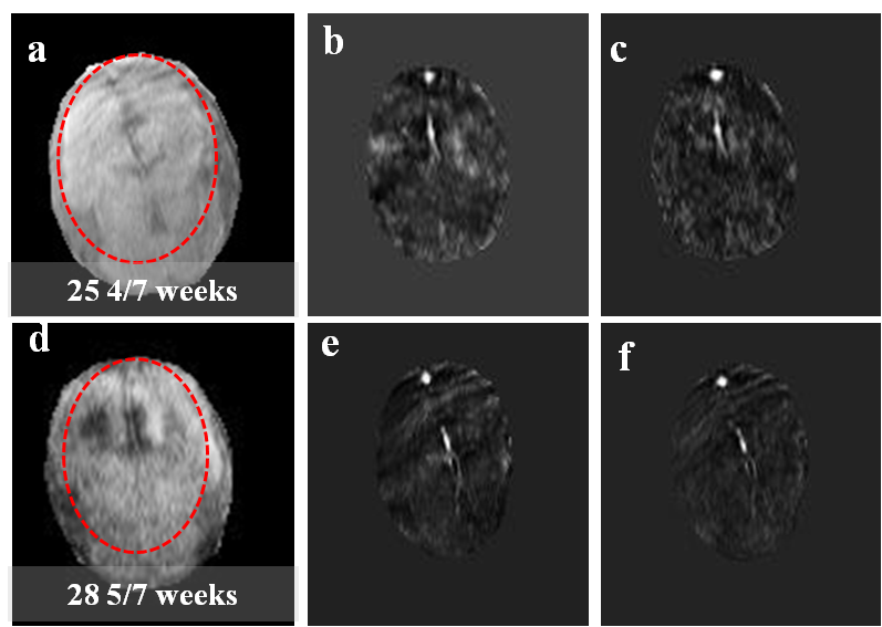
A modified 2D STrategically Acquired Gradient Echo (STAGE)4 sequence was applied on 21 singleton fetuses with enlarged ventricles (Gestational age: mean = 29.6 ± 4.9 weeks) at 1.5 Tesla Siemens Aera System scanner with 8-channel body coil and a spine coil. MRI parameters are given in Table 1. A total of 19 susceptibility weighted imaging (SWI) images encompassing the entire fetal brain were acquired within 1 minute. The QSM maps were estimated as described elsewhere3 are as follows: 1) The raw phase of each image was corrected for global phase offset and then subjected to a background field removal using homodyne filtering (16x16); 3) A 3D brain mask was manually extracted from the high-pass filtered phase images; 4) Minimum of 5 continuous slices of the resultant phase data was then used as input to the iterative, geometry constraint based thresholded k-space division algorithm for generating QSM; 5) A region-of-interest (ROI) containing at least 10 voxels across SSS was drawn inside the SSS in the QSM images, and the mean and standard deviation of magnetic susceptibility of the SSS (∆χv) was measured; 7) The putative SvO2 in the SSS was quantified assuming ∆χdo = 4* π * 0.27 ppm5 and gestational age dependent fetal hematocrit values from the literature6; 8) Finally, the mean and standard error of ∆χv and SvO2 were obtained across all the fetuses.Results
The average SvO2 in the entire cohort, second and third trimester fetuses were, 68.2% ± 5.1%, 72.5%± 5.9% and 66.1% ± 4.7%, respectively. In addition, a decreasing trend in the SvO2 across gestation was found (slope= -0.8 ± 0.4; p=0.05) however the trend was not found to be statistically significant (Figure-2). We found no statistically significant difference in the SvO2 between second and third trimester fetuses (p=0.13).Discussion
We found a decreasing trend in the SvO2 across the gestation ages however, this trend was not found to be statistically significant. This decreasing trend and average SvO2 in the entire fetal cohort obtained were in close agreement with the healthy fetal literature3,7. This finding may be indicative of adaptation of fetal metabolism in order to maintain the cerebral oxygenation by changing other parameters such as cerebral blood flow which is known to increase to compensate the lack of oxygen. Therefore, in future combined estimation of cerebral SvO2 and blood flow might provide us a complete understanding of the fetal cerebral metabolism in the enlarge ventricle anomalies. STAGE MRI successfully provided rapid fetal image acquisition and mitigated the fetal motion artifacts significantly. This technique holds a tremendous significance in the future in human fetal imaging. It can become a potential tool for rapidly obtaining multi-parametric and quantitative information that may further assist in clinical diagnosis.Conclusion
We report for the first time the estimation of cerebral SvO2 in human fetuses with enlarged ventricles using QSM. This approach is robust and might be beneficial in accessing and monitoring the metabolic status of the fetuses in various clinical conditions.Acknowledgements
No acknowledgement found.References
1. Gaffney, G., et al. "Clinical associations of prenatal ischaemic white matter injury." Archives of Disease in Childhood-Fetal and Neonatal Edition 70.2 (1994): F101-F106.
2. Back, S.A. "Perinatal white matter injury: the changing spectrum of pathology and emerging insights into pathogenetic mechanisms." Mental retardation and developmental disabilities research reviews 12.2 (2006): 129-140.
3. Yadav, B. K., et al. "Quantitative susceptibility mapping in the human fetus to measure blood oxygenation in the superior sagittal sinus." European radiology (2018): 1-10.
4. Chen, Y., et al. "STrategically Acquired Gradient Echo (STAGE) imaging, part I: Creating enhanced T1 contrast and standardized susceptibility weighted imaging and quantitative susceptibility mapping." Magnetic resonance imaging 46 (2018): 130-139.
5. Tang, J., et al., Improving susceptibility mapping using a threshold‐based k‐space/image domain iterative reconstruction approach. Magnetic resonance in medicine, 2013. 69(5): p. 1396-1407.
6. Weisskoff, R.M. and S. Kiihne, MRI susceptometry: Image‐based measurement of absolute susceptibility of MR contrast agents and human blood. Magnetic Resonance in Medicine, 1992. 24(2): p. 375-383.
7. Yadav, B.K., et al. "Imaging putative foetal cerebral blood oxygenation using susceptibility weighted imaging (SWI)." European radiology (2018): 1-7.
Figures