1635
Hyperpolarized 129Xe dissolved-phase MR detects physiological changes in human lungs after low-dose inhaled lipopolysaccharide challengeAgilo L Kern1,2, Filip Klimes1,2, Andreas Voskrebenzev1,2, Marcel Gutberlet1,2, Heike Biller2,3, Julius Renne1,2, Olaf Holz2,3, Frank Wacker1,2, Jens M Hohlfeld2,3,4, and Jens Vogel-Claussen1,2
1Institute of Diagnostic and Interventional Radiology, Hannover Medical School, Hannover, Germany, 2Biomedical Research in Endstage and Obstructive Lung Disease Hannover (BREATH), Member of the German Center for Lung Research (DZL), Hannover, Germany, 3Department of Clinical Airway Research, Fraunhofer Institute for Toxicology and Experimental Medicine, Hannover, Germany, 4Department of Respiratory Medicine, Hannover Medical School, Hannover, Germany
Synopsis
Low-dose inhalation of lipopolysaccharide (LPS) provides a disease model in humans for development of anti-inflammatory drugs but sensitive methods for assessment of the inflammatory response to LPS are lacking. The feasibility of hyperpolarized 129Xe dissolved-phase imaging and chemical shift saturation recovery (CSSR) was investigated in this setting. The ratio of 129Xe in red blood cells and in tissue/plasma was found to decrease and the capillary transit time derived from CSSR was found to increase after LPS inhalation. These effects are attributed to pulmonary edema and vasodilation. In conclusion, hyperpolarized 129Xe MR is sensitive even for low-dose LPS challenges in humans.
Introduction
Chronic obstructive pulmonary disease (COPD) poses a major burden to the world population, being the 4th leading cause of death in 2015.1 Progression of COPD is associated with neutrophilic airway inflammation and targeted anti-inflammatory drugs would be a promising candidate to prevent disease worsening.2 Unfortunately, such drugs are still lacking. Provocation by lipopolysaccharide (LPS) elicits a neutrophilic inflammatory response in human lungs and is thought to be a safe disease model for drug development of anti-inflammatory drugs. Previously, dissolved-phase MRI/MR spectroscopy of 129Xe in tissue-plasma (TP) and red blood cells (RBC) has been shown to be feasible for disease monitoring after segmental provocation of human lungs by bronchoscopy.3 However, the invasiveness of repeated bronchoscopies limits the use of this technique. Low-dose inhalation challenges of the whole lung using LPS represent a less invasive alternative but the detection of its functional effects remains challenging.4 Therefore, the aim of this work was to test the feasibility of hyperpolarized 129Xe dissolved-phase MRI and MR spectroscopy for monitoring the response of human lungs after low-dose LPS inhalation.Methods
This study was approved by the institutional review board. 13 healthy
volunteers were included, out of which 10 completed the study
protocol. Hyperpolarized 129Xe MRI was performed at baseline and 6
hours after provocation. For provocation, the subjects inhaled 2µg LPS as
described previously.4
Imaging was
performed at 1.5T (Avanto, Siemens Healthineers, Erlangen, Germany) using a 129Xe
birdcage transmit coil and 16-channel receive array (Rapid Biomedical, Rimpar,
Germany). Hyperpolarization of 129Xe was achieved using a commercially
available polarizer (9810, Polarean, Durham, NC, USA).
The imaging
protocol consisted of a 129Xe dissolved-phase imaging (DPI) scan5 and chemical shift saturation recovery
spectroscopy (CSSR6) during separate breathholds. Starting from
residual volume, subjects inhaled 1L of hyperpolarized 129Xe for DPI
and a variable amount of air to achieve a lung inflation of 1/3 forced vital
capacity. For CSSR, after inhalation of a mixture of 0.5L 129Xe and
high-purity nitrogen, subjects further inhaled room air to fully inflate their
lungs.
DPI data
was separated into RBC and TP phase using hierarchical IDEAL7 and whole-lung mean values for the
ratios TP-GP (GP, gas phase), RBC-GP and RBC-TP were computed. 129Xe
gas uptake data to the TP phase from CSSR was fitted using the Patz model and
physiological parameters were extracted.8 High-resolution spectra of the dissolved phase
acquired directly after DPI were fitted with two complex Lorentzians for
lineshape analysis.Results
For DPI, the whole-lung ratios RBC-TP (Wilcoxon’s signed rank test, p = 0.004) and RBC-GP (p = 0.020) were significantly reduced after provocation compared to baseline. There also was a trend for elevated TP-GP ratio, which was not significant (p = 0.074). See Figure 1 for DPI results. For CSSR, the capillary transit time was significantly increased after provocation (p = 0.020), corresponding to slower blood flow. There was a trend for elevated surface-to-volume ratio (p = 0.084). No significant change was observed for septal wall thickness (p = 0.193). CSSR results are summarized in Figure 2. Spectroscopic lineshape analysis exhibited a significant increase in T2* of the TP phase (p = 0.004) and a significant reduction in RBC chemical shift (p = 0.023), Figure 3.Discussion
This study demonstrates the sensitivity of hyperpolarized 129Xe dissolved-phase MRI and MR spectroscopy for the response of human lungs to low-dose inhalation challenges with LPS. The reduction of the ratios RBC-TP and RBC-GP in DPI is likely due to the presence of increased fluid content of the lungs due to edema after LPS inhalation, leading to diffusion restriction. This is consistent with the slight increase in T2* of TP, which is expected to occur in lungs with reduced air fraction.9 The increase in capillary transit time from CSSR can be explained by vasodilation due to LPS-induced inflammation, leading to reduced blood flow velocity. An increased fluid content of the lung would imply an increase of the alveolar wall thickness parameter and of the surface-to-volume ratio obtained from the TP phase. However, only a trend towards elevated surface-to-volume ratio was observed, likely due to the relatively small sample size. The apparent reduction of chemical shift of the RBC phase could be a sign for a reduced oxygenation of the RBCs due to a diffusion limitation of oxygen.10 However, given that the mean difference observed in RBC Larmor frequency is only ~0.2ppm, further research is necessary to validate this finding.Conclusion
Hyperpolarized 129Xe dissolved-phase MR is highly sensitive for the effects of inhaled LPS challenge in humans and is therefore suitable as a research tool for drug development.Acknowledgements
No acknowledgement found.References
- World Health Organization. WHO methods and data sources for country ‐ level causes of death 2000-2015. http://www.who.int/healthinfo/global_burden_disease/GlobalCOD_method_2000_2015.pdf. Published 2016. Accessed July 12, 2017.
- Singh D, Siew L, Christensen J, et al. Oral and inhaled p38 MAPK inhibitors: Effects on inhaled LPS challenge in healthy subjects. Eur J Clin Pharmacol. 2015;71(10):1175-1184. doi:10.1007/s00228-015-1920-1.
- Kern AL, Biller H, Klimes F, et al. Hyperpolarized 129Xe MR functional imaging to monitor the response of the human lungs after segmental lipopolysaccharide challenge. In: Proc Intl Soc Mag Reson Med 26. ; 2018:2436.
- Janssen O, Schaumann F, Holz O, et al. Low-dose endotoxin inhalation in healthy volunteers - a challenge model for early clinical drug development. BMC Pulm Med. 2013;13(1):1. doi:10.1186/1471-2466-13-19.
- Qing K, Mugler JP, Altes TA, et al. Assessment of lung function in asthma and COPD using hyperpolarized 129 Xe chemical shift saturation recovery spectroscopy and dissolved-phase MRI. NMR Biomed. 2014;27(12):1490-1501. doi:10.1002/nbm.3179.
- Kern AL, Gutberlet M, Qing K, et al. Regional investigation of lung function and microstructure parameters by localized 129 Xe chemical shift saturation recovery and dissolved-phase imaging: A reproducibility study. Magn Reson Med. September 2018. doi:10.1002/mrm.27407.
- Tsao J, Jiang Y. Hierarchical IDEAL: Fast, robust, and multiresolution separation of multiple chemical species from multiple echo times. Magn Reson Med. 2013;70(1):155-159. doi:10.1002/mrm.24441.
- Patz S, Muradyan I, Hrovat MI, et al. Diffusion of hyperpolarized 129 Xe in the lung: a simplified model of 129 Xe septal uptake and experimental results. New J Phys. 2011;13(1):015009. doi:10.1088/1367-2630/13/1/015009.
- Christman RA, Ailion DC, Case TA, et al. Comparison of calculated and experimental NMR spectral broadening for lung tissue. Magn Reson Med. 1996;35(1):6-13. doi:10.1002/mrm.1910350103.
- Wolber J, Cherubini A, Leach MO, Bifone A. Hyperpolarized 129Xe NMR as a probe for blood oxygenation. Magn Reson Med. 2000;43(4):491-496. doi:10.1002/(SICI)1522-2594(200004)43:4<491::AID-MRM1>3.0.CO;2-6.
Figures
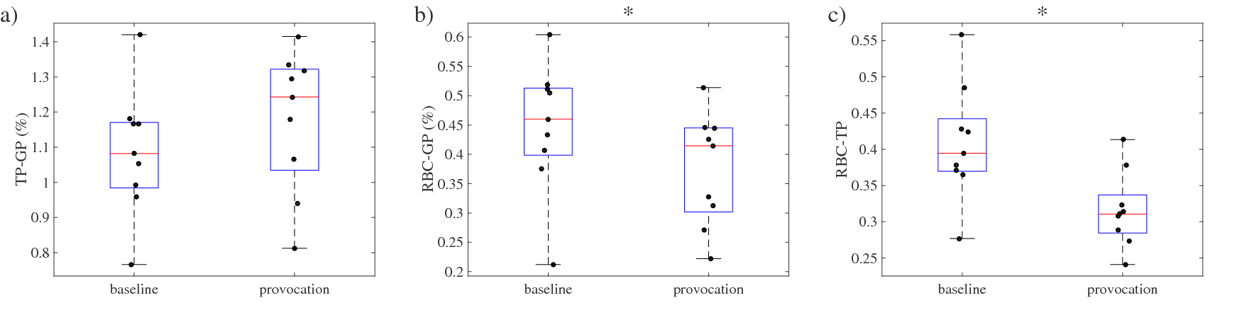
Whole lung mean of a) TP-GP ratio, b) RBC-GP ratio
and c) RBC-TP ratio obtained from DPI in all subjects at baseline and after inhalation
of LPS. Boxes show group median, 25th and 75th percentile,
whiskers extend to most extreme data points. Asterisks
mark statistical significance.
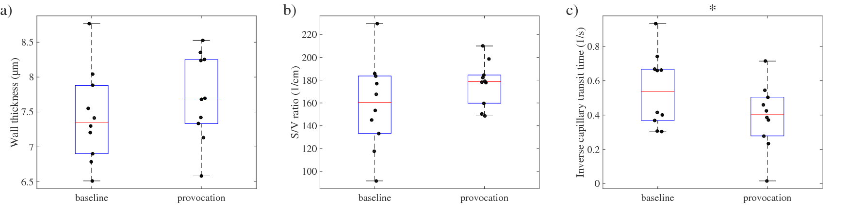
Results of Patz model fits to CSSR uptake curves
for a) septal wall thickness, b) surface-to-volume ratio and c) inverse
capillary transit time corresponding to blood flow velocity. A significant reduction in blood flow velocity is
observed.
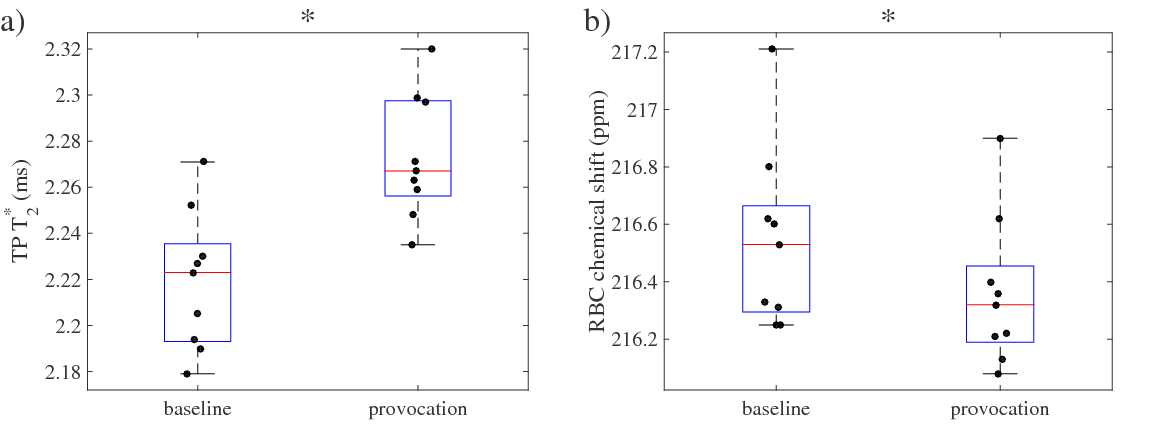
Results from spectral lineshape analysis for a)
transverse relaxation time T2* of TP resonance and b)
chemical shift of RBC resonance.