1634
Noise Reduction in Prostate Single-Shot DW-EPI utilizing Compressed SENSE Framework1Philips Japan, Tokyo, Japan, 2Kumamoto University Hospital, Kumamoto, Japan, 3Philips Healthcare, Best, Netherlands, 4Graduate School of Medical Sciences, Kumamoto University, Kumamoto, Japan, 5Philips Healthcare, Tokyo, Japan
Synopsis
DWI is a key component of the prostate MRI examination, but current prostate DWI images have limited resolution. Small-FOV DWI with SENSE often suffers from increased noise artifacts. We attempt to utilize a combination of parallel imaging and compressed sensing technique (C-SENSE) framework for reducing the noise artifacts in single-shot DW-EPI images (EPI with C-SENSE: EPICS). EPICS clearly reduces noise-like artifacts and significantly improves the accuracy and robustness of ADC values in small FOV high b-value prostate DWI compared with conventional SENSE DW-EPI, without any penalty for scan parameters.
PURPOSE
Diffusion-weighted imaging (DWI), including an apparent diffusion coefficient (ADC) map and high b-value images, is a key component of the prostate multi-parametric MRI exam1,2. Currently, typical clinical prostate DWI images have limited resolution and a larger field-of-view (FOV) compared to other imaging, such as T2 weighted images and contrast-enhanced images3. In fact, small-FOV DWI with SENSE (sensitivity encoding) often suffers from increased noise-like artifacts on the center of the images due to the high geometry factor when large acceleration factors are used with respect to the coil geometry characteristics4,5.
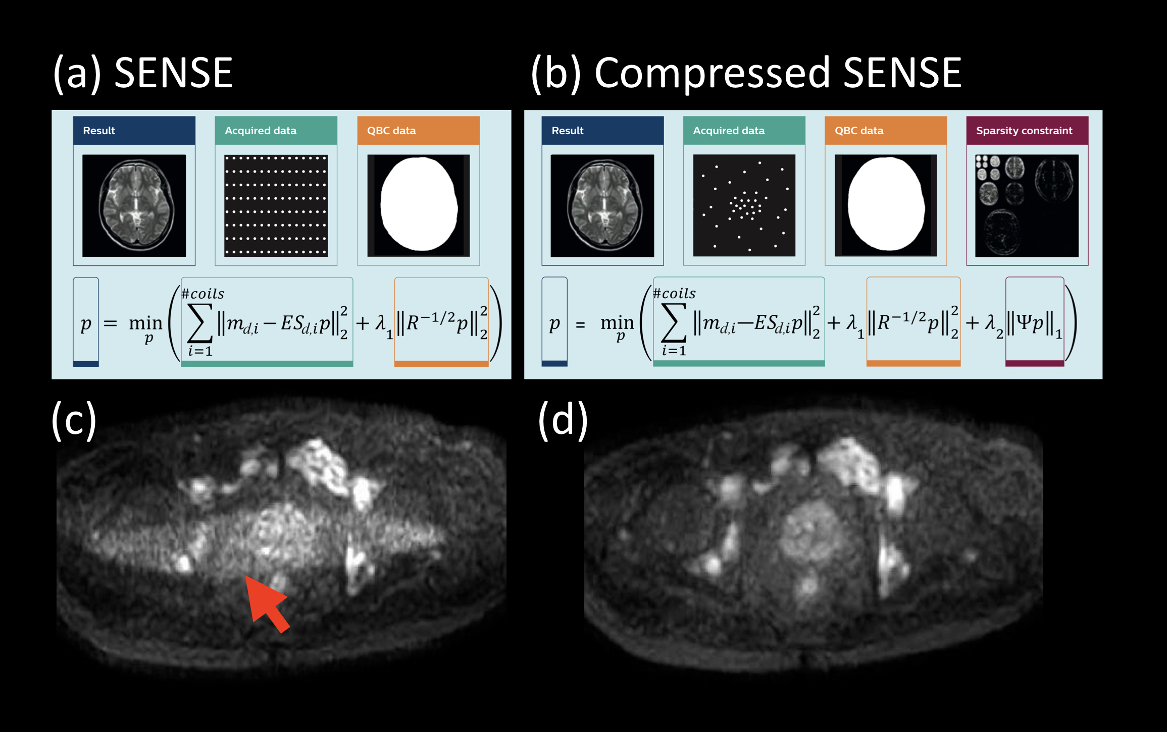
Recently, a combination of parallel imaging and compressed sensing technique (Compressed SENSE, C-SENSE) (Fig1) has been developed to accelerate the acquisition time without increasing the image artifacts6. In C-SENSE, the SENSE-based balanced sampling pattern is automatically optimized using a variable density incoherently under-sampled scheme. For C-SENSE reconstruction, an iterative L1-minimization optimization is used to achieve an optimal balance between noise reduction and data consistency for CS6,7. Although this technique is basically used for shortening acquisition time, it sometimes dramatically reduces the severe noise artifacts, particularly in turbo spin-echo (TSE)8-based DWI (Fig1.c and d.). Now, we hypothesize that the C-SENSE reconstruction can similarly improve image quality for EPI based DWI, without further optimization of EPI sampling scheme. It has been shown before that wavelet-based denoising is an effective tool for image quality improvement in high b-value DWI images9,10. In this work, it is integrated with SENSE parallel imaging in an iterative implementation.
We attempt to utilize the C-SENSE framework for reducing the noise artifacts in single-shot DW-EPI images (EPI with C-SENSE: EPICS). The purpose of this study is to demonstrate the clinical usefulness of EPICS in small-FOV prostate DWI with b-value of 1400 s/mm2 in volunteers and patients, compared to conventional SENSE-DWI.
METHODS
A total of ten volunteers and two patients were examined on 3.0T systems (Ingenia CX, Philips Healthcare). The study was approved by the local IRB, and written informed consent was obtained from all subjects.
EPICS is based on single-shot DW-EPI acquisition. We did not modify its sampling pattern and applied the C-SENSE framework for reconstruction.
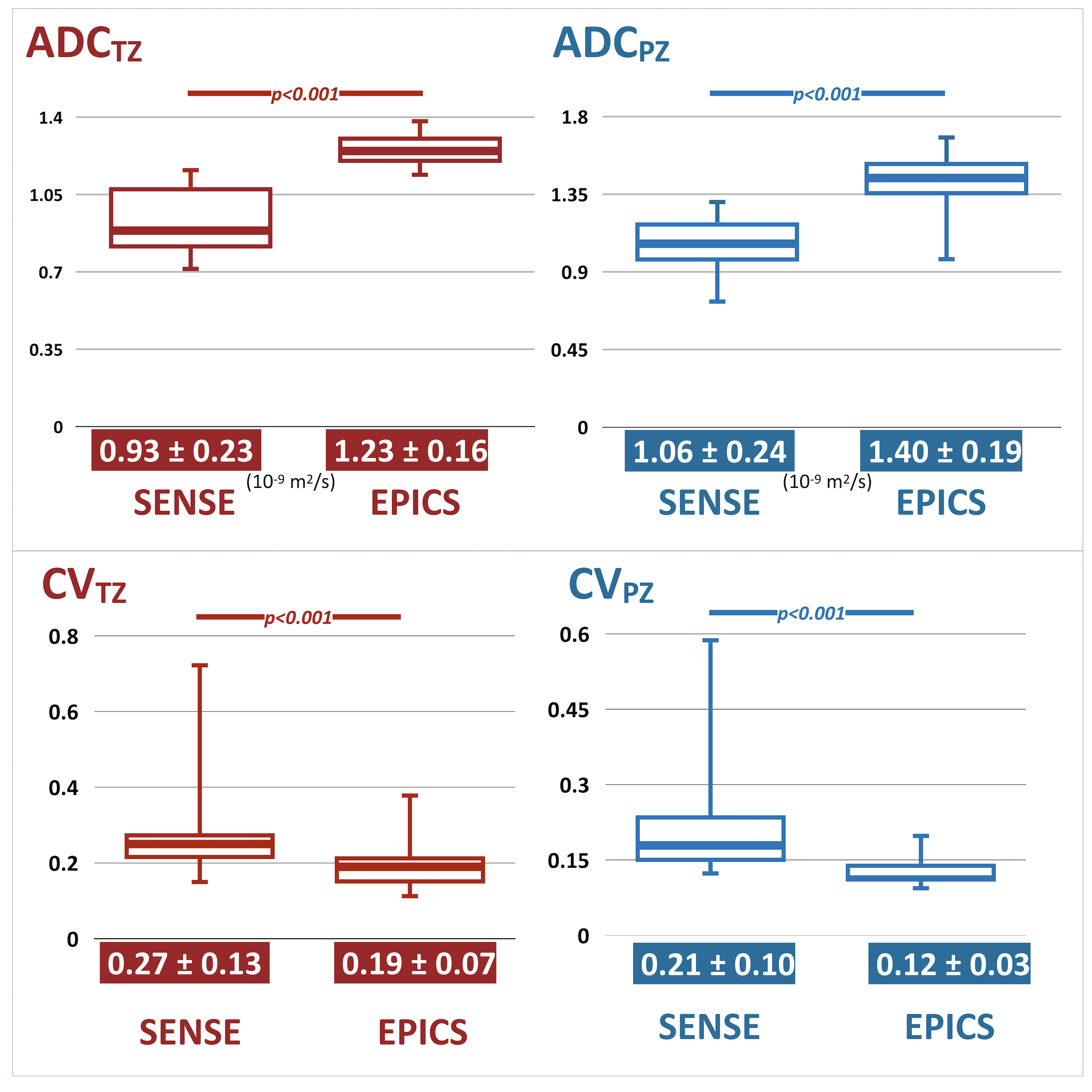
EPICS-DWI images were compared with conventional SENSE images for image quality, especially for the reduction of image noise. To demonstrate the improvement of ADC values by the effect of EPICS noise reduction, we compared ADC values with a SENSE scan with exactly the same scan parameters. ROIs were placed on left and right transition zones (TZ), and left and right peripheral zones (PZ). The average ADCs of TZ and PZ were used for comparison between SENSE and EPICS. We also compared with the values reported in literature11. Furthermore, we calculated the Coefficient of Variation (CV) of ADC values inside the ROIs in respective volunteers as follows: CV = SDADC / AverageADC, to evaluate the consistency of ADC values. Respective ADC and CV values were assessed by using paired t-test.
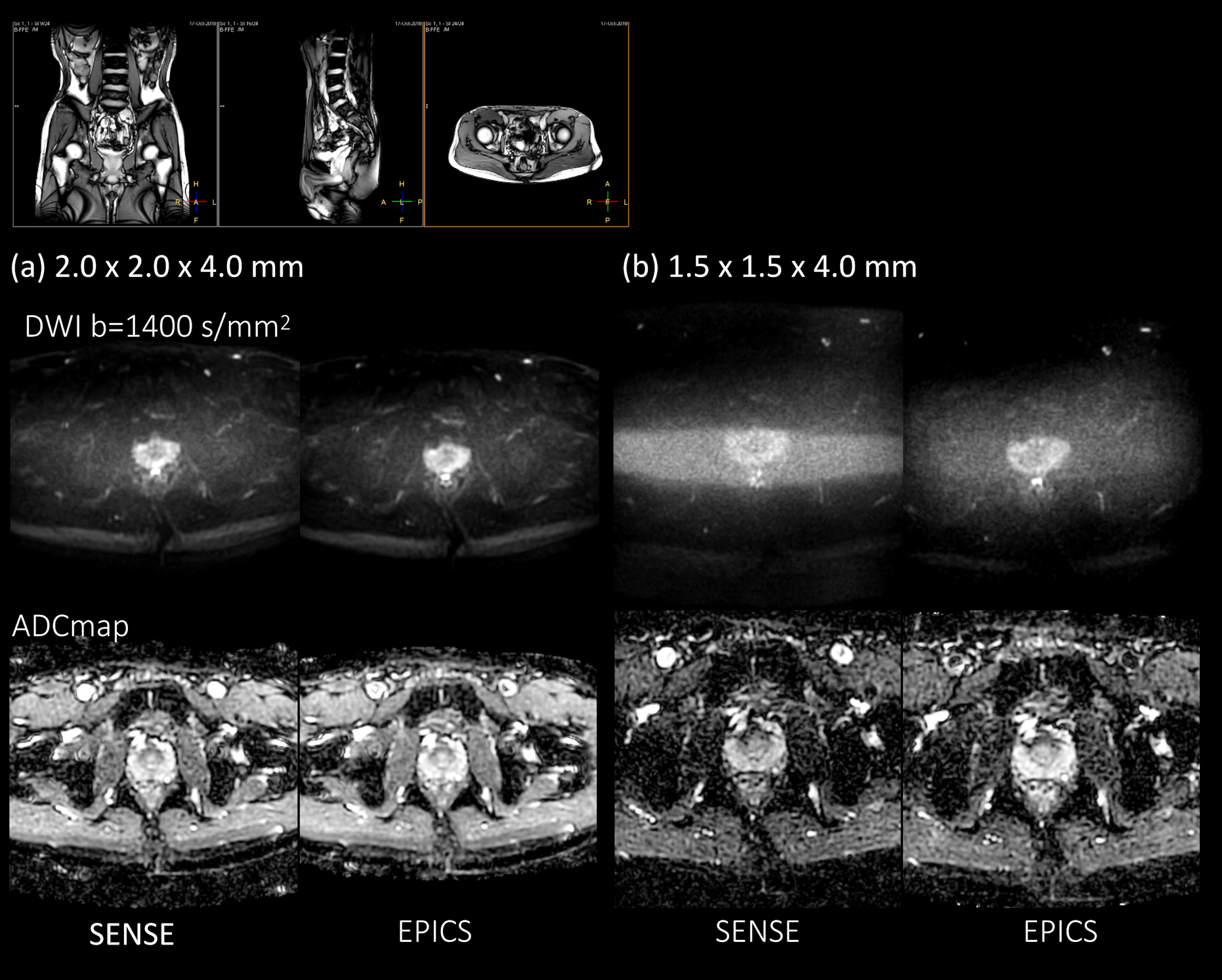
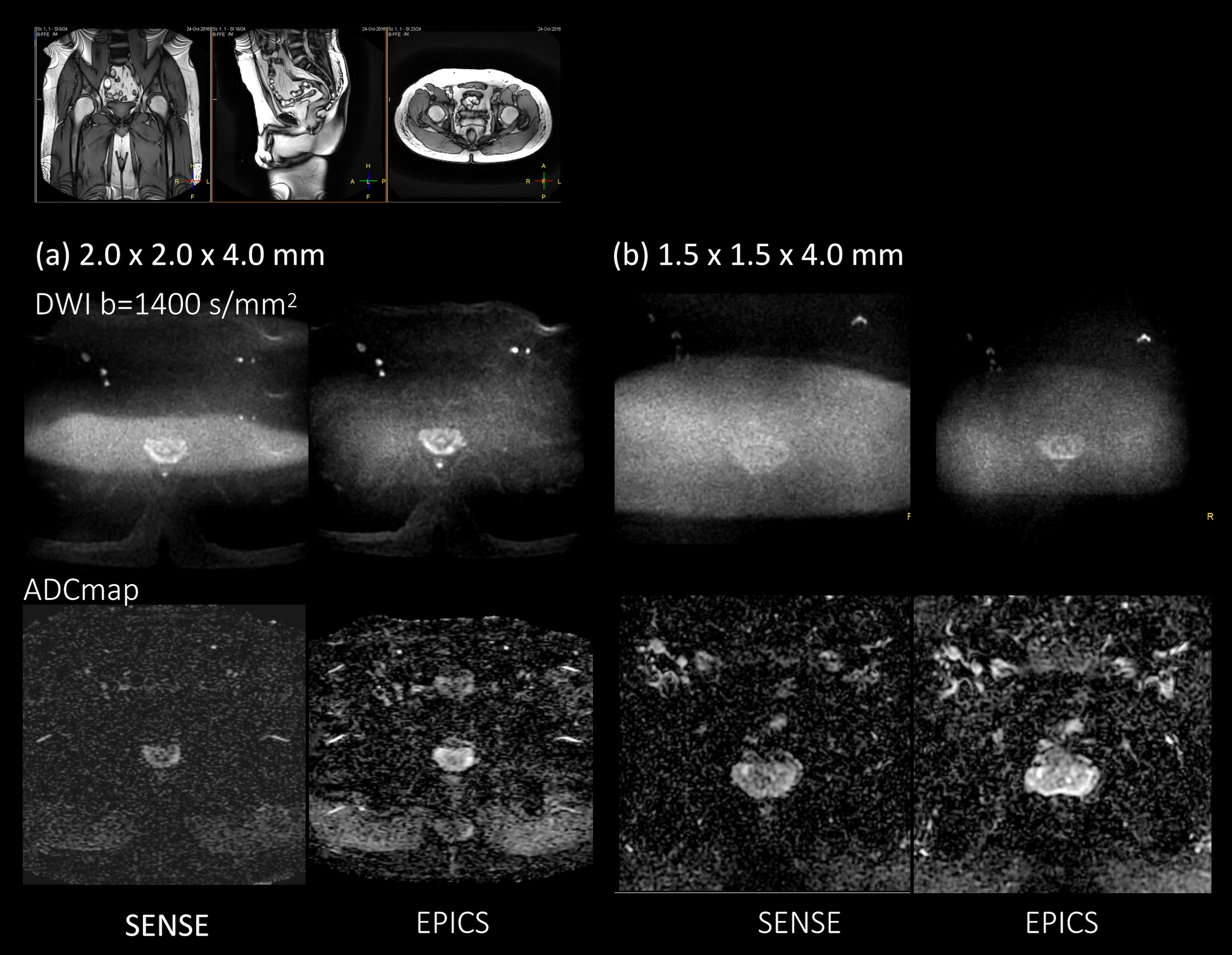
Imaging parameters for DWI were based on PI-RADS consideration1: voxel size=2*2*4mm3, FOV=260*260mm, 20slices, b-value=0 and 1400s/mm2, TR=4000ms, TE=78ms, SENSE/C-SENSE acceleration factor=2.5, and total acquisition time=4min00s. Moreover, we also acquired and compared the smaller FOV/higher spatial resolution protocol as follows: voxel size=1.5*1.5*4mm3, FOV=200*200mm, 20slices, b-value=0 and 1400s/mm2, TR=46000ms, TE=78ms, SENSE/C-SENSE factor=2.5, and scan duration=4min50s. SENSE/C-SENSE factor of 2.5 in anterior-posterior direction was chosen to exemplify the high g-factor behavior in this study even though normally an acceleration factor of 2 is advised in anterior-posterior direction.
RESULTS and DISCUSSION
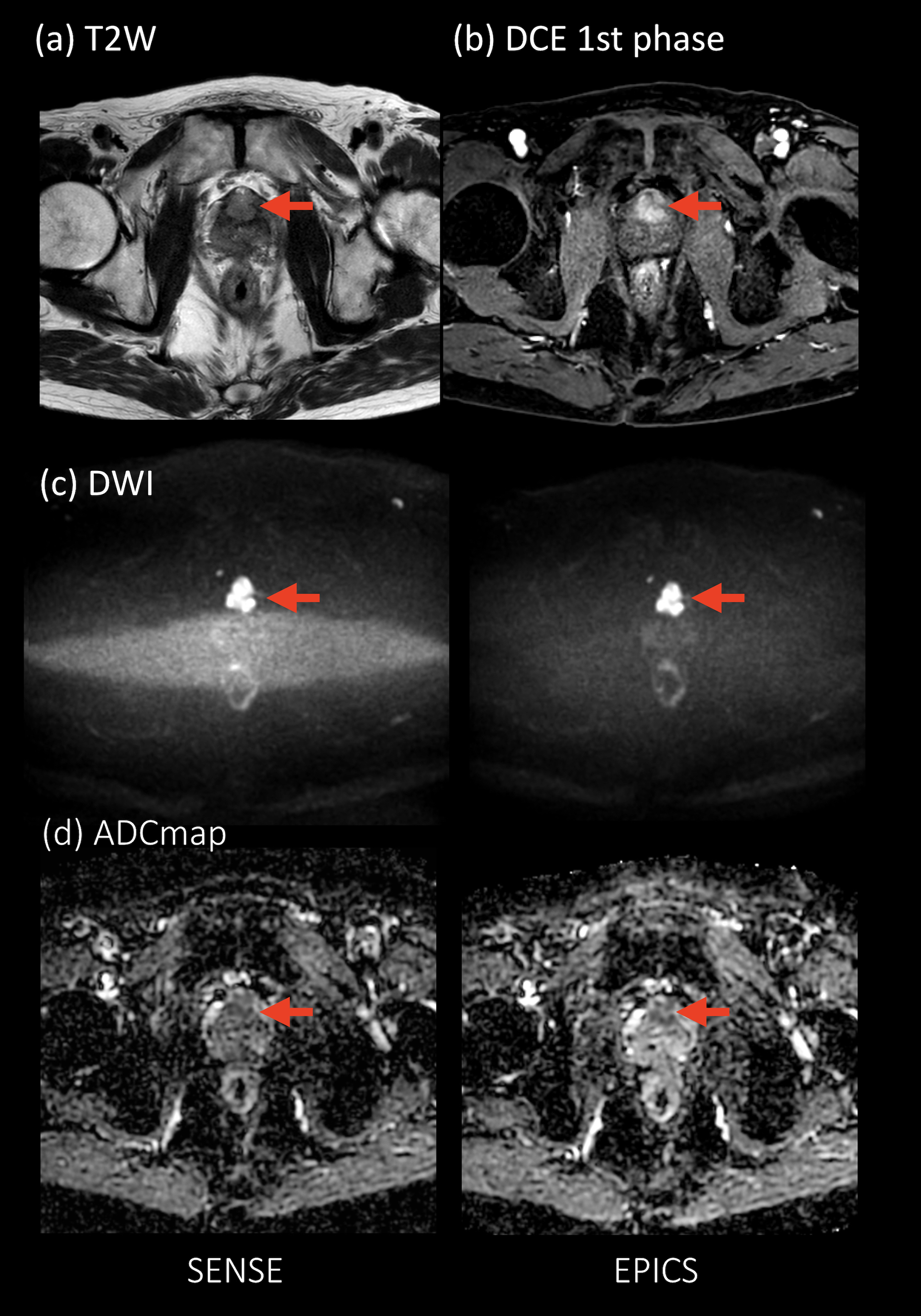
Fig.2 and 3 show the representative images of a relatively skinny volunteer and an obese volunteer. EPICS clearly reduced the noise which exists in the center of the SENSE-DWI images. Fig.4 shows the ADC and CV values of TZ and PZ. The ADC values with EPICS were close to the values reported in literature11 whereas the values with SENSE were lower, most likely due to the presence of severe noise over the prostate. Furthermore, CV values of EPICS were significantly lower than those of SENSE. It indicated that EPICS can provide more accurate ADC values with high reproducibility and robustness. Fig.5 shows the representative images in a patient with prostate cancer. In SENSE-DWI, “noise-band” artifacts obscured accurate interpretation of cancer localization. In contrast, EPICS cleaned up the noise artifact and clearly improved the lesion conspicuity, not only in DWI images but also in ADC map. Although further clinical investigation is needed, EPICS might be clinically useful in assessment of prostate cancer in more detail.CONCLUSION
EPICS clearly reduces noise-like artifacts and significantly improves the accuracy and robustness of ADC values in small-FOV high b-value prostate DWI compared with conventional SENSE-DW-EPI, without any penalty for scan parameters. This technique may be helpful to further assess prostate cancer pathology.Acknowledgements
No acknowledgement found.References
1. Weinreb JC, et al. PI-RADS prostate imaging–reporting and data system: 2015, version 2. European Urology 2016;69:16-40.
2. Tan CH, et al. Diffusion-weighted MRI in the detection of prostate cancer: meta-analysis. AJR Am J Roentgenol. 2012;199:822-9.
3. Medved M, et al. High-resolution diffusion-weighted imaging of the prostate. AJR Am J Roentgenol. 2014;203:85-90.
4. Patricia N, et al. Parallel Imaging Artifacts in Body Magnetic Resonance Imaging. Can Assoc Radiol J. 2009;60: 91–98.
5. Yanasak NE, et al. MR imaging artifacts and parallel imaging techniques with calibration scanning: a new twist on old problems. Radiographics. 2014;34:532-48.
6. Geerts-Ossevoort L, et al. Compressed SENSE Speed done right. Every time. The Netherlands: Philips Healthcare; 2018 Jan. Report No: 4522 991 31821. https://www.philips.de/content/dam/b2bhc/de/resourcecatalog/landingpages/ingeniaelition/White_Paper_Compressed_SENSE-opt.pdf
7. Bratke G, et al. Accelerated MRI of the Lumbar Spine Using Compressed Sensing: Quality and Efficiency. J Magn Reson Imaging. 2018. doi: 10.1002/jmri.26526.
8. Akamine Y, et al. Reduced distortion in prostate DWI by using split echo type TSE-DWI (SPLICE) with MultiVane acquisition. Proc Intl Soc Mag Reson Med. 2018;26:0344.
9. Wirestam R, et al. Denoising of Complex MRI Data by Wavelet-Domain Filtering: Application to High-b-Value Diffusion-Weighted Imaging. Magn Reson Med. 2006;56:1114-20.
10. Yang X, et al. A wavelet multiscale denoising algorithm for magnetic resonance (MR) images. Meas Sci Technol. 2011;22:025803.
11. Wang X, et al. High-b-value diffusion-weighted MRI for the detection of prostate cancer at 3 T. Clin Radiol. 2014;69:1165-70.
Figures