1628
Assessment of the prostate cancer with HyperCube T2-weighted imagingMotoyuki Katayama1, Takayuki Masui1, Kazuma Terauchi1, Mitsuteru Tsuchiya1, Masako Sasaki1, Kenshi Katayama1, Takahiro Yamada1, and Mitsuharu Miyoshi2
1Seirei Hamamatsu General Hospital, Hamamatsu, Japan, 2GE Healthcare Lapan, Hino, Japan
Synopsis
We compare delineation of prostate cancers in HyperCube T2WI with those in conventional T2WI using PIRADS. HyperCube 3D T2WI can provide useful information about prostate cancer, and contribute to the PI-RADS.
Introduction
T2-weighted imaging (T2WI) has played an important role in evaluation of the prostate cancer. Especially, on Prostate Imaging and Reporting and Data System (PI-RADS): version 2.0, T2WI is the main diagnostic tool of the cancer in the transition zone. However, without using the endorectal coil, it was difficult to obtain the high spatial resolution T2WI in a shorter acquisition time. HyperCube (GE Healthcare) expands the capabilities of 3D imaging with partially selective excitation technique similar to FOCUS, and enables to significantly reduce scan times and eliminate artifacts with very selective saturation pulse by reducing the phase field of view. In addition, HyperSense (GE Healthcare), which is a combination of ARC and compressed sensing, enables to obtain the images within shorter acquisition time. The purpose of this study is to compare delineation of prostate cancers in HyperCube T2WI with those in conventional T2WI using PI-RADS version 2.0.Materials and Methods
Patients: 29 male patients, suspected of having cancers in the prostate gland and confirmed with biopsy and/or surgery, who underwent MRI on a 3T unit (Signa Pioneer, GE Healthcare) with a 32 channel phased array coil, were included in this study. Age: 67.6 years old, ranged from 49-82 years old. MR imaging: The spatial resolution of HyperCube T2 was as follows; TR/TE: 2002/ 94 msec, FOV: 26x13cm, Matrix: 416*224, section thickness: 1.6 mm, the number of excitation: 2, total acquisition time: 169 seconds, and that of conventional T2WI was as follows; TR/TE: 4000/102 msec, FOV: 20*20 cm, Matrix 384*256, section thickness: 4 mm, the number of excitation: 4, total actuation time: 164 seconds, fast recovery technique applied, respectively. Evaluations Qualitative analyses (overall image quality, distortion, blurring, motion artifact, and delineation of prostate tissue, respectively) of each imaging were performed with 5-point scale. As a qualitative analysis, the contrasts of the signal intensities (SI) of the tumor against those of the prostate tissue adjacent to the tumor in each imaging were calculated. In order to avoid the selection bias, we evaluated the largest one lesion on each the patient. The scores on PI-RADS version 2.0 assessment of T2WI were applied to the tumor detection on each imaging.Results
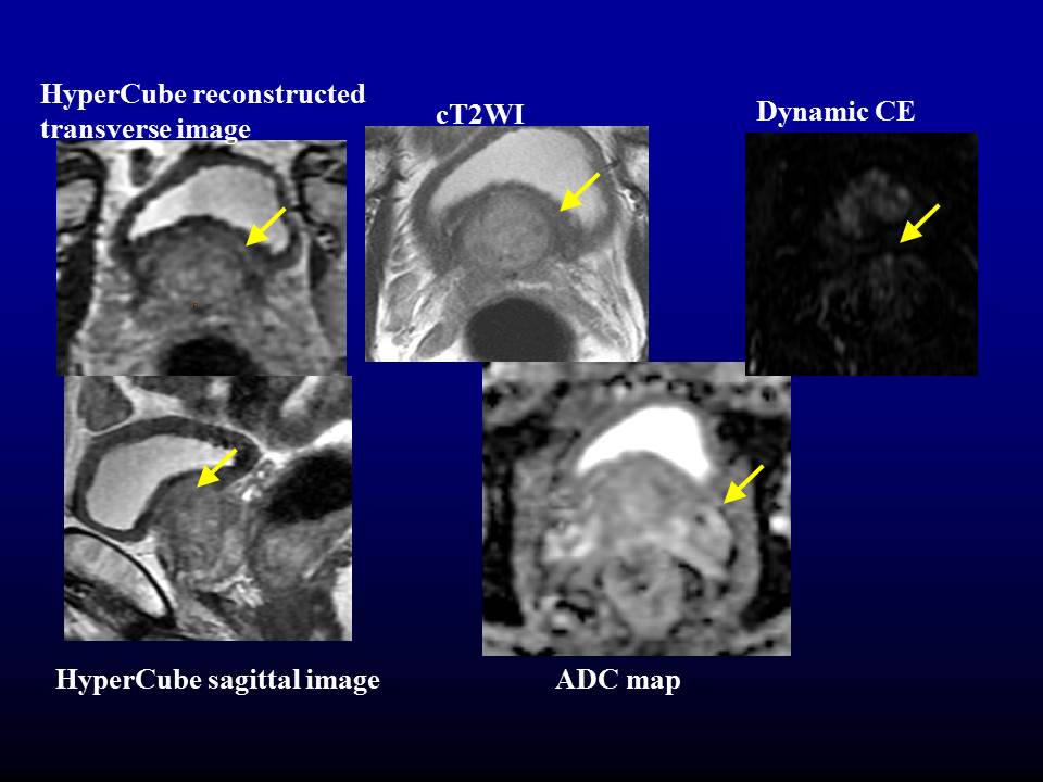
Summary of the patients’ data were as follows; Clinical stage of the prostate cancer Negative: n = 5 ≦T2: n = 22, T3: n = 1 (T3a) T4 : n = 1 (invasion of urinary bladder), transition zone: n = 6, peripheral zone: n = 18, maximum Gleason score (GS) of the tumor; GS 6: n = 5, GS 7 (3+4): n = 8, GS 7 (4+3): n = 6, GS 8≦ n = 5. The total prostectomy were performed in 14 patients. With the qualitative analyses of each imaging, almost all images showed good image quality (figure 1). The contrast of tumor against the prostate tissue adjacent to the tumor in Hypercube was superior to that in cT2WI (figure 2). The weighted Kappa score of 0.95 shows higher reproducibility of each imaging on PI-RADS version 2.0(figure 3). Figure 4 and 5 show the images of HyperCube T2WI and conventional T2WI, respectively.Discussion
We demonstrated the usefulness of HyperCube T2WI for evaluation of prostate cancer. In order to evaluate with PI-RADS version 2.0 strictly, the high special resolution images (section thickness: equal or less than 4mm) were required, although it takes longer time to acquire the above images without endorectal coil. Generally, 3D T2-weighted images may be particularly useful for visualizing detailed anatomy and distinguishing between genuine lesions and partial volume averaging effects. However, the soft tissue contrast is not identical and in some cases may be inferior to that seen on 2D T2-weighted images. With HyperCube T2-weighted imaging technique, we can acquire such as high spatial and high contrast images within a few minutes. Our study had some limitations. First, our study was retrospectively performed in the single center. The selection bias of the patients might occur. Second, the sample size of our study was small. Third, the sampling error of needle biopsy might happen to our patients. Forth, the total prostectomy were performed in only 14 of 29 patients. Finally, the location and size of each tumor was not confirmed exactly.Conclusion
HyperCube 3D T2WI can provide useful information about prostate cancer, and contribute to the PI-RADS.Acknowledgements
No acknowledgement found.References
No reference found.Figures
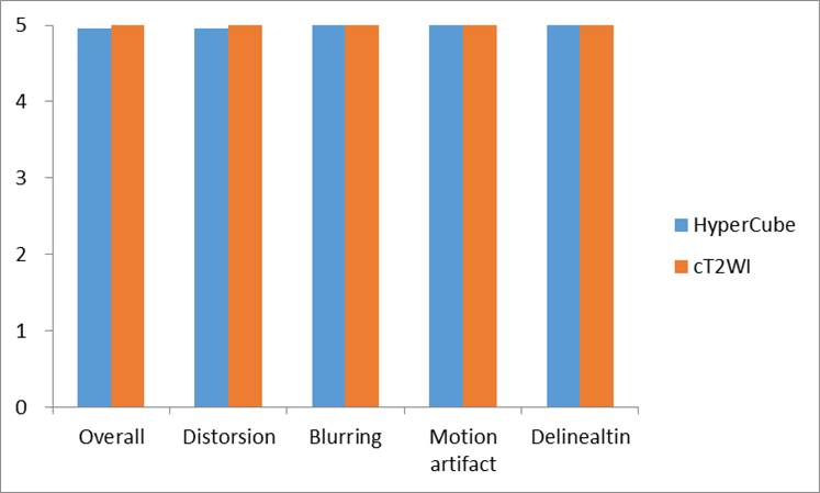
Qualitative
analyses
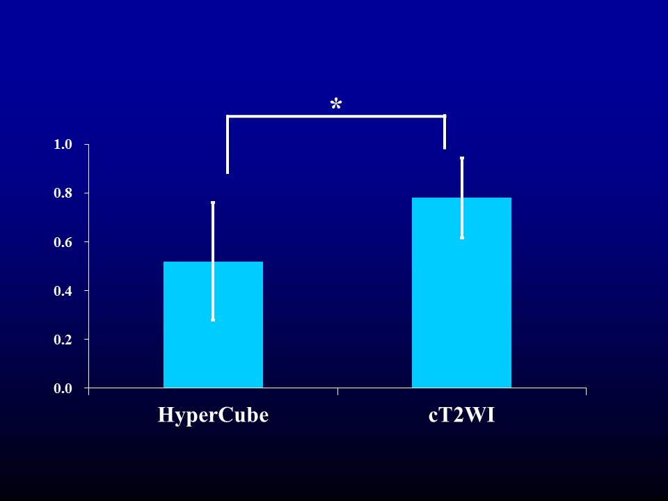
The contrast
of tumor against the adjacent prostate tissue
adjacent to the tumor.
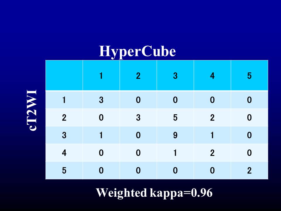
PI-RADS
scores: weighted Kappa analysis
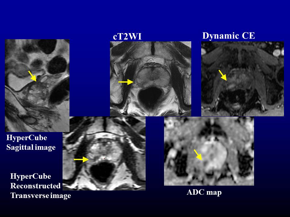
74 year-old male with GS =
3+4 tumor in the right mid-PZpm
area
75 year-old male with GS = 4+3
in the left base TZa area