1611
Test-retest repeatability of ADC measurements using MUSE: Evaluation in phantoms and prostate1Memorial Sloan Kettering Cancer Center, New York, NY, United States
Synopsis
In this study, we evaluated the repeatability of multiplexed sensitivity-encoding (MUSE) DW-EPI apparent diffusion coefficient measurements (ADC) in phantom and prostate images. High quality test-retest prostate and phantom ADC maps obtained from phantom and volunteer studies measured values using MUSE (2-4 interleaves) were within 1.4-4.7% (phantom) and 3.2-14.7% (prostate) of one another. In comparison, test-retest repeatability results for standard single-shot EPI (acceleration factor=2) were 4.5-9.4% (phantom) and 15.2-19.2% (prostate). MUSE images exhibit reduced geometric distortion.
PURPOSE:
The aims of this study were to investigate the clinical feasibility and to report test-retest repeatability of ADC measurements using multiplexed sensitivity-encoding (MUSE) DW-EPI in phantom and prostate images. Test-retest ADC maps generated with MUSE (2-4 interleaves) were compared with maps generated separately using standard single-shot EPI (ss-EPI, acceleration factor=2) in a phantom with known ADC values and in the prostate.INTRODUCTION:
The MUSE acquisition and reconstruction platform was developed for brain imaging to enable DW-MR images to be acquired with reduced geometric distortion using an interleaved acquisition. Nonlinear shot-to-shot phase variations are corrected without the need for navigator echoes though (1). Recently, MUSE was evaluated for brain radiotherapy applications (2). To the best of our knowledge, no prior study evaluated the repeatability of MUSE in phantom and prostate studies. In the study, we present preliminary repeatably results for ADC measurements using MUSE.METHODS:
All MRI studies were performed on a 3T unit (GE DV26 platform). A body coil was used for excitation. For signal reception, a cardiac eight-channel phased-array coil was used. For all experiments, DW images were obtained using both conventional ss-EPI (SENSE=2) and MUSE, twice. Table 1 summarizes the variants. All images were acquired with FOV=28cm, 4mm thickness; matrix, 11 slices, b=0 (4 NEX) and 800 s/mm2 (16 NEX), identical axial coverage.
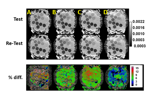
Experiments: For our phantom study, we used a phantom developed in accordance with the Quantitative Imaging Biomarkers Alliance (QIBA) guidelines. The phantom consists of 13 vials (20 mL) with 6 difference concentrations (0%, 10%, 20%, 30%, 40%, 50%) of polymer polyvinylpyrrolidone (PVP) in aqueous solution submerged in an ice water bath which serves to maintain temperature at 0°C. The same protocol was used to image the prostate.
Data Analysis: In the QIBA phantom and prostate studies, regions of interest (ROI) were drawn over regions of each cross-section of the vials as well as the peripheral zone (PZ) and transition zone (TZ) of the prostate. Mean ADC value in the ROI were calculated using all diffusion acquisitions. The repeatability was assessed as 100×|ADCdifference|/ADCmean. The within-subject coefficient of variation (wCV) is given by wCV=100×wSD/mean where wSD is the within-subject standard deviation (wSD) (3).
RESULTS:
Test-retest results for the phantom and prostate are shown in Figs. 1 and 2, respectively. For ss-EPI, the repeatability range was 4.5-9.4%. In comparison, for MUSE, the range of values were 1.7-3% (sequence B), 1.6-4.7% (sequence C), and 1.4-3% (sequence D). For prostate, the repeatability using ss-EPI was 19.2% for PZ and 15.15% for TZ. In comparison, the values using the MUSE sequences B-D were 14.7%, 7.2%, and 8% for PZ, and 7.8%, 3.3% and 4% respectively for TZ (Fig. 3). The wCV values for the QIBA phantom were comparable across sequences, but for prostate, the values were 34.9% and 20.5% in the PZ and TZ respectively, using ss-EPI. In comparison, using sequences B-D the values were 20.5-26.9% for PZ and 13.7-18% for TZ (Fig. 4).DISCUSSION:
The MUSE sequence reduces geometric distortion due to susceptibility effects. Our preliminary results suggest that the estimated ADC values are repeatable in test-retest experiments where the imaging object (phantom, human) is not intentionally displaced during repeat measurements. Due to the greater sensitivity of ss-EPI to air in the rectum as compared to multi-shot techniques, it is expected that MUSE would demonstrate greater repeatability to fluctuations within the body (such as air or bowl motion) which can result in susceptibility. Our preliminary test-retest results are consistent with this expectation.CONCLUSION:
Based on our preliminary results, we conclude that the MUSE platform enables repeatable DW imaging of the prostate. Additional studies are needed to evaluate its clinical utility in detection and staging of cancer.Acknowledgements
No acknowledgement found.References
1. Chen NK, Guidon A, Chang HC, Song AW. A robust multi-shot scan strategy for high-resolution diffusion weighted MRI enabled by multiplexed sensitivity-encoding (MUSE). NeuroImage 2013;72:41-47.
2. Chen X, Zhang Y, Cao Y, et al. A feasible study on using multiplexed sensitivity-encoding to reduce geometric distortion in diffusion-weighted echo planar imaging. Magnetic resonance imaging 2018;54:153-159.
3. Newitt DC, Zhang Z, Gibbs JE, et al. Test-retest repeatability and reproducibility of ADC measures by breast DWI: Results from the ACRIN 6698 trial. Journal of magnetic resonance imaging : JMRI 2018.
Figures