1610
Two exploratory radiomics segmentation algorithms in T2-weighted imaging analysis for predicting apical positive surgical margins of prostate cancer: A pilot study1Peking University First Hospital, Beijing, China
Synopsis
SynopsisThis retrospective study aims to compare the two different segmentation algorithms of radiomics analysis for predicting the apical surgical margin (SM) status before radical prostatectomy (RP). Preoperative prostate MR scans were performed for 76 enrolled patients and T2-weighted images was assessed by a radiomics model, using two different delineating methods: including the surrounding tissues of the targeted lesions or not. Finally 152 bilateral surgical margins’ status (training dataset: n = 110, testing dataset: n = 38) were evaluated. The result demonstrated the segmentation algorithms were comparable, of which the new method might reduce the delineation work for future radiomics research.
Introduction
Prediction of positive surgical margins preoperatively is a promising topic in radiomics research, which may offer a reliable pretreatment information to the surgeons in order to select appropriate patient candidate or surgical approaches1,2. However, the delineation of possible inked margins of the pathological section is time-consuming and the standard has not been developed and adopted.
Objective
To compare the predictive performance of two different radiomics segmentation algorithms in apical surgical margin status at radical prostatectomy (RP) based on preoperative T2-weighted MR imaging.
Methods
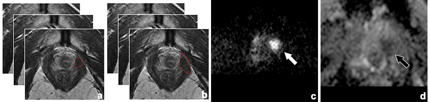
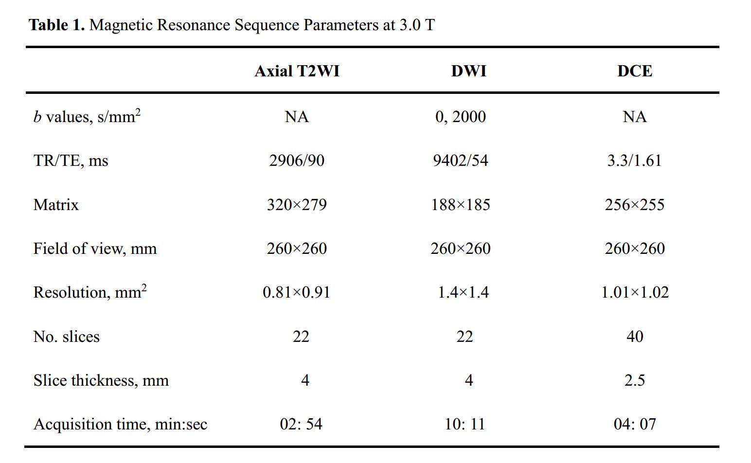
This IRB-approved retrospective study included 76 patients with prostate cancer (PCa) undergoing multi-parametric MRI before prostatectomy from January 2014 to December 2017 (Table 1). Radiomics analysis based on derived T2-weighted imaging features was performed on 152 bilateral surgical margins confirmed via pathological section, of which there were 59 positive apical surgical margins (PSM) and 51 negative surgical margins in the training cohort, and 11 PSM and 29 negative margins in the testing cohort. Patients were allocated to training and testing cohorts on the basis of the surgical procedures. The radiomics analysis pipeline concerning volume selection and segmentation, features selection, data integration, data mining and interpreting the predictive results. For the first step, two different delineating algorithms were conducted: (1) segmentations only included tissue within the confines of the targeted lesions evaluated as PI-RADS 4-5 by experienced radiologists, (2) the algorithm involving the targeted lesion as well as the possible ink-stained tissues outside the suspected tumor region (Fig. 1). Specificity, sensitively and areas under the curves (AUC) of the radiomics model were calculated and compared in terms of the status of apical surgical margin status. Histopathological sections results was considered as the reference standard.Results
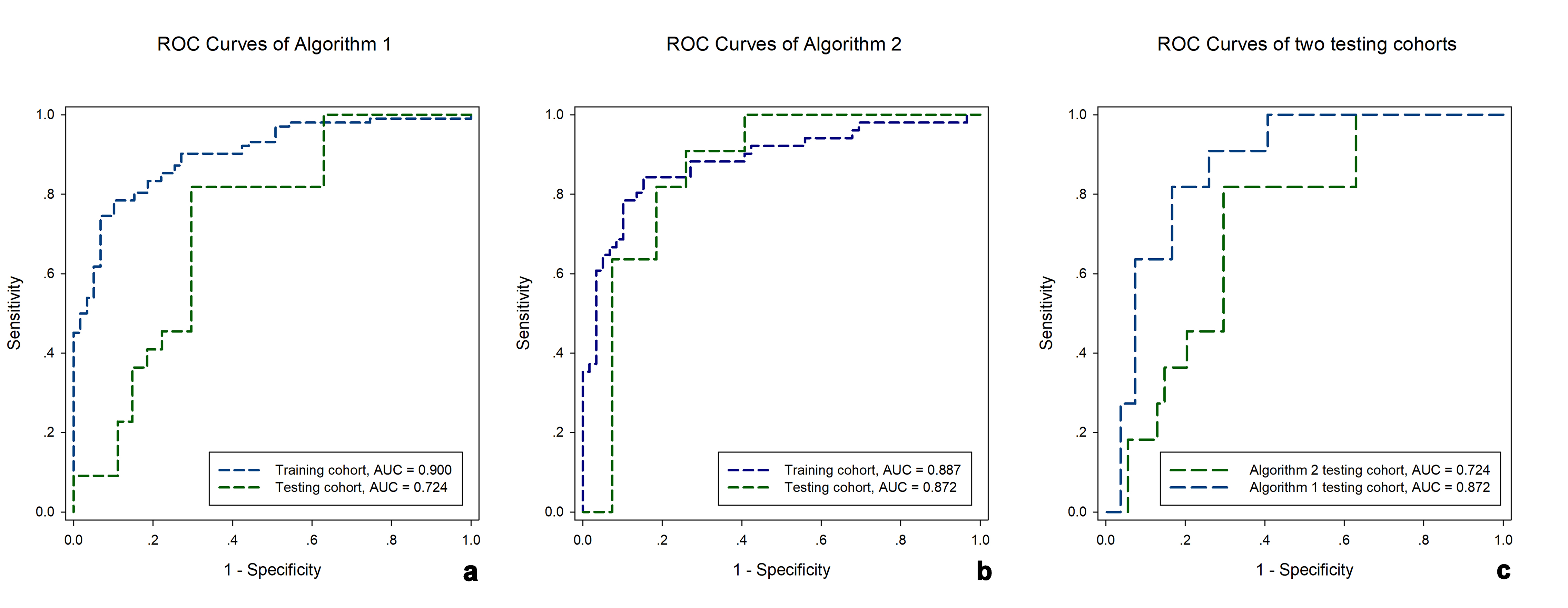
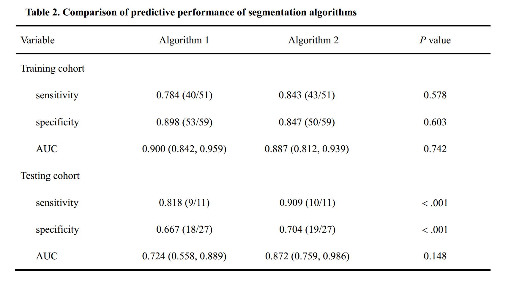
Table 2 summarized the predictive performance of segmentation algorithms. In all, for the testing cohort, the area under the curves (AUC) showed no difference between the two training cohorts (0.900 vs. 0.887, P = 0.742) (Fig. 2), and no significant difference occurred concerning the sensitivity (0.784 vs. 0.843, P = 0.578) and specificity (0.898 vs 0.847, P = 0.603). For the testing cohort, although the set of algorithms 2 demonstrate a better predictive performance with respect to the sensitivity and specificity (0.909 vs. 0.818, and 0.704 vs. 0.667, all P < 0.001), the AUCs showed no difference between the algorithms.s analysis.Discussion
In the current pilot study, although diagnostic performance (AUCs, sensitivity and specificity) did not show a significant difference between the two segmentation methods, a tendency of superiority of the new segmentation algorithm was obvious in terms of area under the curves, sensitivity and specificity, demonstrating that such exploratory delineation methods is at least equally appropriate for predicting the apical surgical margins of PCa, as compared with the conventional delineation way which always performed in the targeted tumor volume itself in the pipeline of the radiomics analysis.Admittedly, the relative small sample size in the radiomics model may be a main influence factor in the current study.Conclusion
In this study, compared to an individual tumor lesion within the surgical margin, we were able to show that the radiomics analysis of the entire prostate tumor which contained tumor itself with the addition of the surrounding possible ink-stained tissues is a valid approach, for the preoperative assessment of apical surgical margins. This exploratory segmentation algorithm might be translated to other tumor entities, which might reduce the delineation work in the workflow of the radiomic analysis.Acknowledgements
No acknowledgement found.References
1. Stoyanova R, Takhar M, Tschudi Y, et al. Prostate cancer radiomics and the promise of radiogenomics. Translational cancer research 2016;5(4):432-447.
2. McEvoy SH, Raeside MC, Chaim J, et al. Preoperative Prostate MRI: A Road Map for Surgery. AJR American journal of roentgenology 2018;211(2):383-391.
Figures