1608
Design of a 12-channel rhesus head coil array for functional MRI at 3 T1Shanghai United Imaging Healthcare, Shanghai, China, 2Lauterbur Imaging Research Center, Shenzhen Institutes of Advanced Technology, Chinese Academy of Sciences, Shenzhen, China, 3Department of Radiology and Biomedical Imaging, University of California San Francisco, San Francisco, CA, United States, 4UCSF/UC Berkeley Joint Graduate Group in Bioengineering, San Francisco, CA, United States
Synopsis
In this work, a
12-channel rhesus head coil array for functional magnetic resonance imaging
(fMRI) was designed, constructed and evaluated by imaging experiments in
phantom studies and in-vivo studies. Compared to a commercially available
12-channel knee coil, the proposed 12-channel rhesus head coil provides
improved performance not only in SNR and parallel imaging capability, but also
in temporal SNR (tSNR) in resting-state fMRI studies.
Introduction
The rhesus monkey is an important animal model used in functional magnetic resonance imaging (fMRI) to understand the brain1. To achieve high-temporal resolution fMRI for rhesus monkey, phase array coils2,3 with high SNR and parallel imaging capability need to be applied. Due to the special brain structure of rhesus monkey, the commercially available received coils are not applicable because of the low SNR and inappropriate coil geometry. In this work, we designed and built a dedicated 12-channel rhesus head coil array for a 3T system (uMR 790, Shanghai United imaging Healthcare, Shanghai, China). The performance of the dedicated 12-channel rhesus head coil, in term of SNR, parallel imaging capability, and temporal SNR in resting-state fMRI studies, was evaluated in comparison with that of a commercially available 12-channel knee coil array.Methods
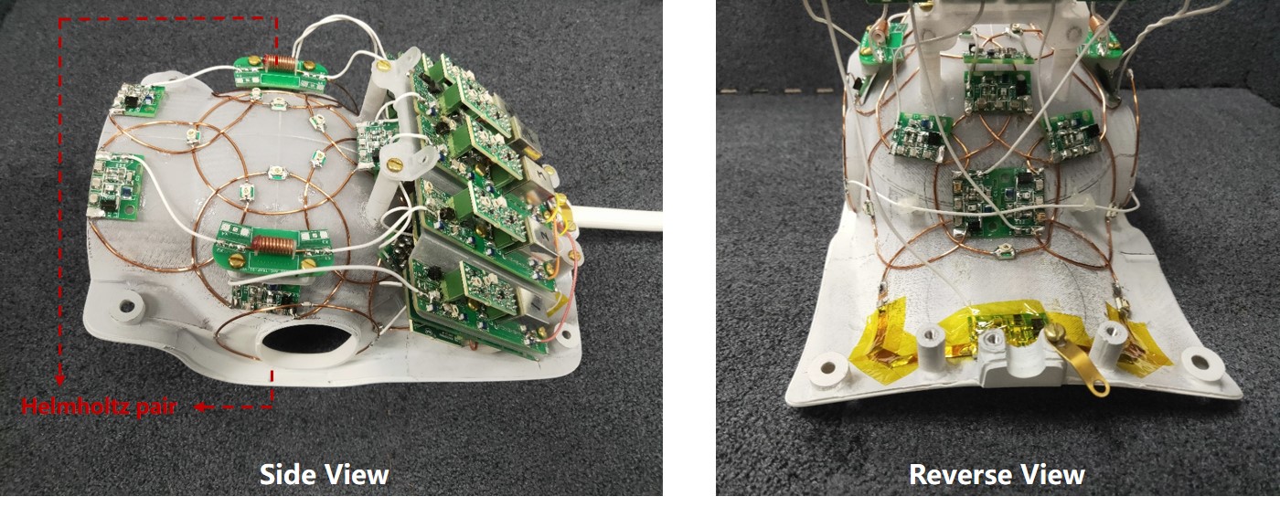
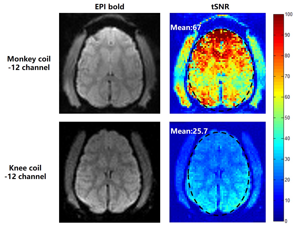
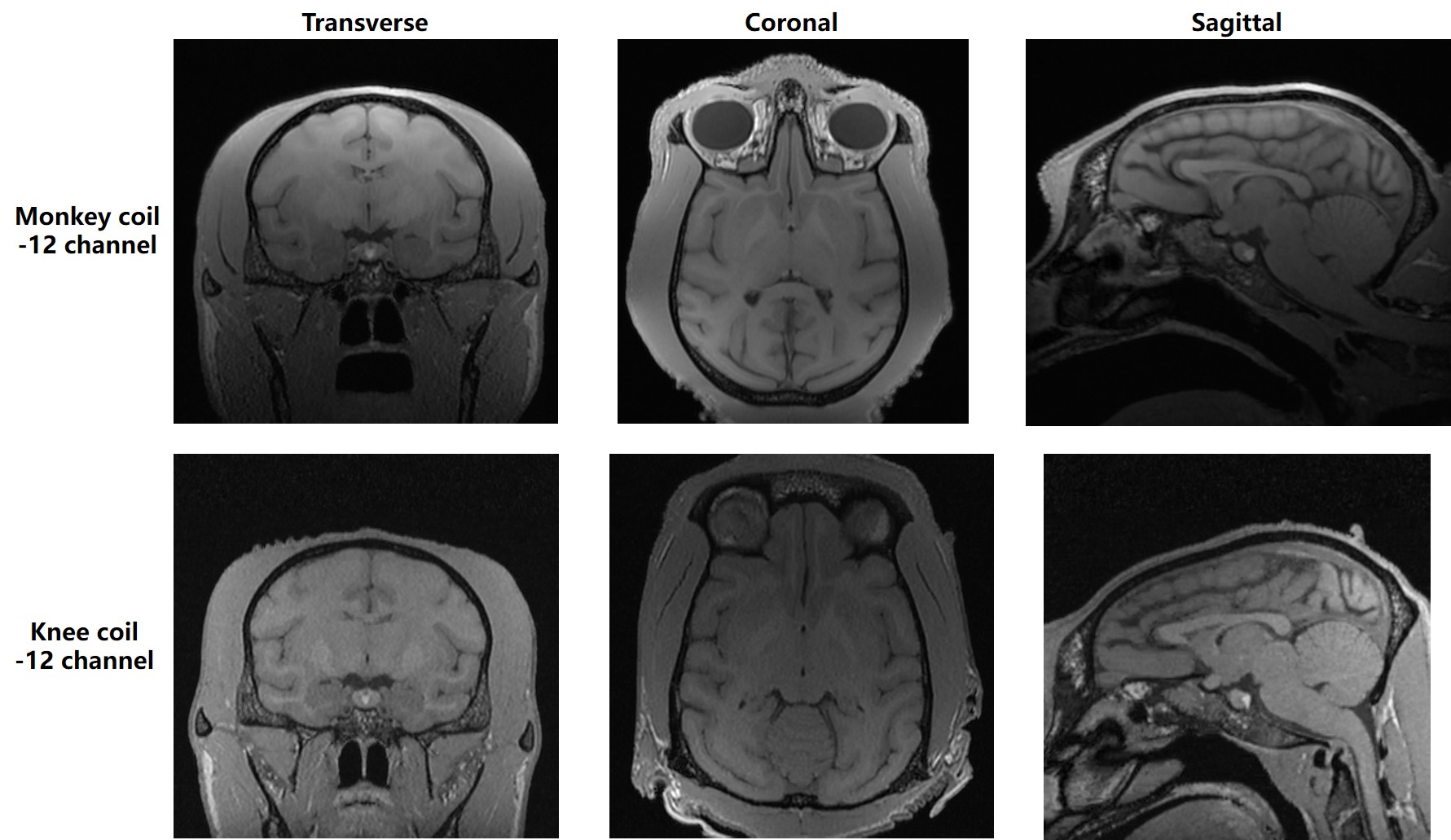
As shown Fig. 1, the 12-chanel rhesus head coil includes two parts: a Helmholtz pair of circle loops in a diameter of 70 mm connected with coaxial cable that were placed on the ear of the rhesus, and the other 11 elements with diameter of 65 mm placed around the head to cover the whole-brain. All the studies was implemented on a 3T MRI scanner (uMR 790, Shanghai United Imaging Healthcare, Shanghai, China) and all experiments of the 12-channel rhesus head coil were performed in comparison with that of a 12-channel commercially available knee coil. Phantom study: A customized sphere phantom with a diameter of 10 cm2 was made to simulate the rhesus head, and was filled with (2.62g NiSO4•6H2O+1.24g NaCl)/1000ml H2O. The phantom signal images were acquired with a 2D GRE sequence (TR/TE=300 ms/15 ms, flip angle=30o, FOV= 120 x 120 mm2, slice thickness=5 mm, Matrix size=256 x 256) and the noise was acquired with the same sequence when flip angle=0o. The signal and noise results can be applied to compute covariance weighted SNR and SENSE g-factor maps4. In-vivo study: For the resting-state fMRI study, a gradient echo BLOD EPI sequence was applied with the parameters as following: TR/TE= 2100/25 ms, flip angle=90o, FOV=96×96 mm2, acquisition matrix =64×64, slices=32, resolution=1.5 × 1.5 × 2 mm3, phase-encoding=H-F, R=2, bandwidth= 2000 Hz/pixel, and repetitions=130. The temporal SNR (tSNR) maps to evaluate the EPI-BOLD image quality were calculated by dividing the mean signal intensity of 130 times by the standard deviation5. The anatomical images were acquired by T1-weighted 3D GRE FSP sequence with following parameters: TR/TE/TI=8.2 ms/2.7 ms/1210 ms, FOV=100x100 mm2, slice thickness=0.5 mm, slices=220, matrix=190x190, isotropic resolution 0.52x0.52x0.5 mm3, flip angle=10o, bandwidth=250 Hz/pixel, number of average=2.Results
Fig. 2 shows the comparison of SNR maps between the 12-channel Monkey coil and the 12-channel knee coil. The mean SNR values in the circle regions, as depicted in the figure, are calculated and shown in the corresponding positions. SNR improvement is obvious in the deep brain regions, which is at least 71 %. Fig. 3 depicts the inverse g-factor maps in the transverse plane with acceleration factors R from 2 to 4 in the Right/Left (R/L) direction and R=2x2, R=3x3. The results indicate that the parallel imaging capability of the 12-channel monkey coil is better than that of the knee coil, particularly at high acceleration factors. Fig. 4 shows the tSNR maps in the resting-state fMRI studies at a coronal plane of Rhesus brain as an example. The mean tSNR values are also evaluated in an ellipse region, as depicted in the maps, from which it can be known that the 12-channel Monkey coil shows a 2.6-fold tSNR improvement. Fig. 5 shows the 3D rhesus monkey brain structural images with a high spatial resolution (0.52x0.52x0.5mm3) acquired by the two coils. Known from the brain images, the proposed monkey coil can achieve a better performance with more clear images in the whole brain region than the knee coil.Conclusion
A custom-designed 12-channel rhesus head coil was designed, constructed and evaluated by phantom studies and in-vivo experiments at 3T. Compared to a commercially available 12-channel knee coil array, the 12-channel Rhesus coil achieves not only better performance in SNR and acceleration ability, but also higher tSNR in resting-state fMRI studies. High resolution 3D images and better image quality for fMRI can be achieved by using the dedicated 12-channel rhesus head coil.Acknowledgements
This work was supported in part by NSFC under Grant No. 61571433, 61801466, 81627901, 81527901; Guangdong Province grants 2014A030312006, and 2014B030301013; Youth Innovation Promotion Association of CAS No. 2017415; city grants JCYJ20170413161314734; NIH U01EB023829, and a Pengcheng Scholar Award.References
1. R. M. Hutchison and S. Everling. “Monkey in the middle: why non-human primates are needed to bridge the gap in resting-state investigations,” Frontiers in Neuroanatomy, vol.6, no.29, pp. 1-19, Jul. 2012.
2. J. Lee, X. Yang, Q. Chen, C. Tie, X. Zhang, H. Zheng, and Y. Li. “An 8 channel Rhesus Head coil for Neuroimaging on 3T,” 2018 ISMRM, Paris, France.
3. J. Lee, X. Yang, Q. Chen, C. Tie, X. Long, N. Li, X. Zhang, H. Zheng, and Y. Li. “Development of an 8channel head array for MRI guided monkey ultrasound stimulation,” 2018 ISMRM, Paris, France.
4. B. Keil, and L. L. Wald. “Massively parallel MRI detector arrays,” Journal of magnetic resonance, vol.229, pp. 75-89, Apr. 2013.
5. M. A. Urbin, H. Xin, C. E. Lang, and A. R. Carter. “Resting-state functional connectivity and its association with multiple domains of upper-extremity function in chronic stroke,” Neurorehabilitation and Neural Repair, vol. 28, no. 8, pp. 761-769, Oct. 2014.
Figures